1,830 posters, 67 videos, 75 audios, 29 topics, 22 sessions, 4,727 authors, 1,697 institutions
ePostersLive by SciGen Technologies S.A. All rights reserved.
RCOG World Congress 2023
June 12 - 14, 2023 | London, UK

EP.0875
A Rare Case of Superior Mesenteric Artery Thrombosis Post Vaginal Delivery
Poster Presenter
Authors
Affiliations
Part of Topic
Obesity
A Rare Case of Superior Mesenteric Artery Thrombosis Post Vaginal Delivery
Abstract:
Introduction: Acute abdominal pain following a vaginal delivery is rare. Various causes of severe acute postpartum abdominal pain include, pelvic infection, thrombophlebitis, ovarian venous thrombosis, acute urinary retention. Mesenteric vessel thrombosis is an uncommon but often fatal form of intestinal ischemia.
Case presentation: Our case describes a 35 years old grand multipara presented three weeks after spontaneous vaginal delivery with severe abdominal pain, abdominal distension, and hematemesis of acute onset. No significant medical history. Past surgical history of bariatric surgery two years back. The patient’s hemodynamic state was unstable and was diagnosed with superior mesenteric artery thrombosis by contrast-enhanced computed tomography and had urgent laparotomy and resection of the bowel segment with the temporary closure of the abdomen. On the next day a second look laparotomy was done later reversal of Roux en Y gastric bypass (gastro-gastric anastomosis, jejuno-jejunal anastomosis, and jejuno-ileal anastomosis), appendectomy was carried out. She was discharged on the 10th day of admission in stable condition with regular follow-up in the surgical outpatient department.
Conclusion: Even after vaginal birth, pregnancy and puerperium raise the risk of thrombosis. Given that this is frequently discovered at a late and irreversible stage, an increased index of suspicion is required for an early diagnosis and rapid therapy to enhance the maternal prognosis. To lower maternal mortality, risk assessments for thromboembolism must be made periodically. To rule out mesenteric artery ischemia in cases of acute abdominal discomfort, abdominal contrast-enhanced computed tomography may be required.
Keywords: Mesenteric vessel thrombosis, bariatric surgery, postpartum, pregnancy, bowel ischemia, thrombosis
Background:
Acute abdominal pain poses a unique diagnostic and therapeutic challenge. Any acute intra-abdominal condition characterized by pain, tenderness, and muscular rigidity, for which emergency surgery should be sought, is referred to as acute abdomen [1]. Acute abdominal pain following an uneventful vaginal delivery is a rare occurrence. Various causes of severe acute postpartum abdominal pain have been mentioned in the literature which includes pelvic infection, thrombophlebitis, ovarian venous thrombosis, and acute urinary retention [2]. Mesenteric artery thrombosis is due to the occlusion of the arterial vascular supply of the intestinal system. It is a severe and potentially fatal illness typically of the superior mesenteric artery (SMA), which provides the primary arterial supply to the small intestine and ascending colon. The occlusion may occur due to in-situ thrombosis of the vessel [3-4]. Pregnancy and puerperium are associated with a shift of coagulation and fibrinolytic system towards hypercoagulability for reducing the risk of bleeding during delivery, but this could result in a higher risk of thrombosis. Apart from pregnancy, higher parity, advanced maternal age, operative delivery, obesity, immobilization, heart disease, history of thrombosis thrombophilia and history of sleeve gastrectomy are other predisposing factors [5-6]. We report a case of 35 years old female who presented with severe abdominal pain on the third week of postpartum following uneventful vaginal delivery.
Case Summary:
We report a case of superior mesenteric vessel thrombosis in a 35-year-old female, Gravida 5 Para 5 three weeks post normal vaginal delivery presented to us with complaints of sudden onset of severe upper abdominal pain, which was progressive in nature, also noted to have abdominal distension associated with dizziness, nausea, and vomiting. Vomitus was blood-tinged. Her delivery was spontaneous and uncomplicated had a smooth postpartum recovery and was discharged in good condition. No significant medical history. Two years back she had Laparoscopic Roux en Y gastric bypass and lost around thirty kilograms. The woman had a body mass index of 27. The patient went to another facility before presenting to our facility and was found to be hypotensive and transferred to our facility for further treatment. On presentation, the patient was looking unwell, hypotensive (89/42mmHg), upper abdominal tenderness, distended, normal bowel sounds, no rigidity, no vaginal bleeding, and legs were soft. A bedside scan revealed intraabdominal fluid collection, meanwhile patient started to deteriorate unable to sit up, hematemesis and systolic blood pressure dropped to 50mmHg.Abdominal tapping was done by a general surgeon and drained serosanguineous fluid collected. Massive blood transfusion protocol was activated, and she received 4 units of RBCs, 1 unit of platelet apheresis and 1 unit of fresh frozen plasma. Despite of resuscitative measure blood pressure responded initially but dropped again.
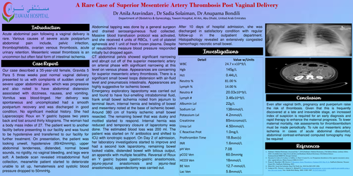
Investigations: (Table : 1) Hemoglobin 14.5g/dL, platelets 309 x109/L, total leukocyte count 24.7 x10⁹/L with neutrophilia, serum creatinine 85 micromol/L , C Reactive Protein 1.0mg/L, metabolic lactic acidosis of 6mmol/L, electrocardiogram normal, Chest Xray unremarkable (Fig 1) , CT abdominal pelvis (Fig 2) showed significant narrowing and abrupt cut off of the superior mesenteric artery on arterial phase with significant narrowing at this level on venous phase. Appearances are concerning for superior mesenteric artery thrombosis. There is a significant small bowel loops distension with air-fluid level and pneumatosis intestinalis. In addition, there is an absence of the mucosal and bowel wall enhancement with significant free fluid in the abdomen and pelvis. Appearances are highly suggestive for ischemic bowel.
Table 1 : Table of investigations :
|
Detail |
Value w/Units |
|
WBC |
24.7 x10^9/L |
|
Hgb |
145 g/L |
|
Hct |
0.44 L/L |
|
Neutro % |
81.00 % |
|
Lymph % |
14.00 % |
|
Neutro # |
20.03 x10^9/L |
|
Lymph # |
3.46 x10^9/L |
|
Albumin Lvl |
22 g/L |
|
Sodium Lvl |
138 mmol/L |
|
Potassium Lvl |
4.2 mmol/L |
|
Creatinine |
85 micromol/L |
|
Urea Lvl |
4.50 mmol/L |
|
C Reactive Prot |
1.0 mg/L |
|
Prothrombin Time |
18.8 sec(s) |
|
INR |
1.6 mmol/L |
|
pH Ven |
7.08 |
|
pCO2 Ven |
60.0 mmHg |
|
HCO3 Ven |
18 mmol/L |
|
BE Ven |
-12.7 mmol/L |
|
Lac Ven |
5.8 mmol/L |
Fig 1: Chest Xray
Fig 2 : CT abdo/pelvis showing There is a significant narrowing and abrupt cut off of the superior mesenteric artery on arterial phase with significant narrowing at this level on venous phase. Appearances are highly concerning for superior mesenteric artery thrombosis.
There is a significant small bowel loop distension with air-fluid level and pneumatosis intestinalis.
Emergency exploratory laparotomy was carried out and found to have foul-smelling intrabdominal fluid, frank small bowel ischemia noted from jejunum to terminal ileum, internal hernia and twisting of bowel and mesentery noted at the base of ischemic bowel. Around 380 cm of frankly ischemic small bowel resected. The remaining bowel that was dusky and mottled started to respond. Internal hernia was reduced and temporary closure of laparotomy was done. The estimated blood loss was 200 ml. The patient was started on IV antibiotics and shifted to ICU with ionotropic support. On Day 2 of admission, her laboratory investigations started to improve and had a second look laparotomy, remaining bowel looked viable, distended bowel with bilious output, an appendix with multiple fecaliths. Reversal of Roux en Y gastric bypass (gastro-gastric anastomosis, jejuno-jejunal anastomosis and jejuno-ileal anastomosis), appendectomy was carried out. She was discharged after 10 days of hospital admission in satisfactory condition with regular follow-up in the outpatient department. Histopathology revealed an extensively congested hemorrhagic necrotic small bowel.
Discussion:
Pregnancy and puerperium increase the risk of thrombosis since they are hypercoagulable states [7]. Coagulability peaks during delivery with placental expulsion due to the release of thromboplastic chemicals, which is a mechanism for reducing maternal blood loss [8].Venous thrombosis, non-occlusive mesenteric ischemia, arterial occlusion by embolism or thrombosis, or both, can cause acute mesenteric ischemia, a serious outcome, and potentially fatal condition [9]. The prognosis typically depends on rapid detection, and arterial thrombosis is typically determined to be the worst condition, followed by non-occlusive and mesenteric embolism. The causes range and include cirrhosis, acute trauma, inflammatory diseases of the hematological disorders, prothrombic states, postoperative states, and decompression sickness[10-11]. Prothrombotic states can occur as a result of oral contraceptive use, pregnancy, neoplasms, antithrombin III deficiency, protein C or S deficiency, factor V Leiden, or obesity. Hematologic disorders include essential thrombocytosis, polycythemia vera, and paroxysmal nocturnal hemoglobinuria. Inflammatory diseases include peritonitis, pancreatitis, intraabdominal infections, diverticulitis, and inflammatory bowel disease[10]. During pregnancy, venous thromboembolism is an uncommon occurrence. The puerperium and pregnancy are both in a hypercoagulable condition. Coagulopathies increase the risk of post-partum thromboembolic events by eightfold[8]. In this case, pregnancy, postpartum, and history of bariatric surgery are all contributing factors in causing mesenteric vessel thrombosis. A Thrombophilia workup was not done on our patient. However, nothing in the patient’s story above matches thrombophilia. Our patient didn’t meet the criteria for postpartum thromboprophylaxis and didn’t receive it as her score was only one (Parity >3).
The presence of one or more risk factors and pain that is out of proportion to physical exam findings should raise clinical suspicion and trigger the required diagnostic workup. Historical findings may include anorexia, nausea, vomiting, and colicky pain often present for more than 48 hours before seeking medical attention. Physical findings may be normal or septic shock due to peritonitis [10].
The diagnosis is not ruled out by normal blood testing, and abnormal results suggest irreversible intestinal ischemia. Late findings are frequently characterized by metabolic (lactic) acidosis and leucocytosis with a left shift. An ileus, intestinal pneumatosis, bowel wall edema with thumbprinting, intraperitoneal air, or portal vein air may all be seen on abdominal radiographs. A normal abdominopelvic ultrasound scan should be followed with an abdominal and pelvic Computed tomography scan [12]. Computed tomography with intravenous contrast is the test of choice and is diagnostic in up to 90% of patients [10, 14-15].
In a stable patient with a small thrombus burden and no peritonitis symptoms, thrombolysis therapy can be initiated. Heparin administered intravenously is the drug of choice for acute management, and warfarin is administered orally for patients whose clot appears stable. Restoring superior mesenteric artery flow and removing non-viable bowel are the mainstays of treatment for a patient with peritonitis. In such circumstances, an embolectomy is also an option [11,16].
There are two possible outcomes following surgical exploration of the lesions: Treatment for localized intestinal necrosis involves resection and prompt restoration of digestive continuity. When the ischemic or infarcted intestine segment is extended, it is challenging to determine the boundaries of resection. To prevent "short bowel syndrome," resection should be effective in every situation. Some teams favor doing a resection followed by a gastrointestinal bypass, along with immediate heparin treatment, followed by a second laparotomy 12 to 24 hours later as done in our case [17].
All patients should be treated with anticoagulation. Additionally, aggressive intravenous hydration, bowel rest, and total parenteral nutrition should be given. The length of anticoagulation is determined by the presence of a prothrombotic state, which may necessitate therapy for life-long. Otherwise, the course of treatment takes 6 to 12 months, just like treating deep vein thrombosis or pulmonary embolism. Acute mesenteric thrombosis typically has a poor prognosis and a mortality rate of 20% to 50% and frequently recurs during the first 30 days. [4, 18].
An effective, multidisciplinary approach for diagnosis and treatment is necessary, comprising physicians, nurses, as well as officials from the lab and radiology. The general surgeon should be consulted early on in the patient's care because there is no specific test to diagnose mesenteric artery thrombosis. Most patients require prompt treatment and resuscitation [19 - 20].
Conclusion:
Even after vaginal birth, pregnancy and puerperium raise the risk of thrombosis. Given that this is frequently discovered at a late and irreversible stage, an increased index of suspicion is required for an early diagnosis and rapid therapy to enhance the maternal prognosis. To lower maternal mortality, risk assessments for thromboembolism must be made periodically. To rule out mesenteric artery ischemia in cases of acute abdominal discomfort, abdominal contrast-enhanced computed tomography may be required.