1,830 posters, 67 videos, 75 audios, 29 topics, 22 sessions, 4,727 authors, 1,697 institutions
ePostersLive by SciGen Technologies S.A. All rights reserved.
RCOG World Congress 2023
June 12 - 14, 2023 | London, UK

EP.0762
HAEMORRHAGIC CYSTIC DEGENERATION OF UTERINE FIBROID PRESENTING AS OVARIAN MALIGNANCY
Poster Presenter
Authors
Affiliations
Part of Topic
Benign Gynaecology
HAEMORRHAGIC CYSTIC DEGENERATION OF UTERINE FIBROID PRESENTING AS OVARIAN MALIGNANCY
Case Report
Objective: Uterine fibroids can have significant effects on the quality of life. More than half of cases are symptomatic, with the peak incidence of symptoms in the 4th decade of life. Uterine fibroids are benign growths of the uterine muscle described histologically as a monoclonal proliferation of smooth cells. Classical clinical presentation includes lower abdominal mass, menorrhagia and pressure symptoms; however, sometimes, they may have an atypical presentation.
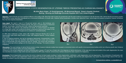
Case report: We report a case of a 45-year-old woman, para 2, Afro-Caribbean, who presented to the A&E following a fainting episode. She gave a 3-month history of abdominal swelling, menorrhagia, worsening pelvic pain, satiety and generalized weakness. On examination, the abdomen was distended and tender. An abdominopelvic CT reported on a large right ovarian cyst containing isodense components layering at the bottom of the cyst, free ]uid in the Pouch of Douglas and no evidence of lymphadenopathy. An MRI study evidenced a bulky uterus with multiple intramural Ibroids and a 25 cm hemorrhagic cystic lesion, likely ovarian in origin, with malignant features. Tumour markers were normal. In view of ongoing symptoms, she had an emergency laparoscopy. Intraoperatively, a large sessile fundal Ibroid mass was identified. Both tubes and ovaries were normal. Four litres of hemorrhagic fluid were drained and sent for cytology. The myoma was morcellated inside an endo-bag without spillage, and the tissue was for histology.
Discussion: The exact aetiology of uterine Ibroid remains unclear. However, molecular studies have revealed a monoclonal nature with specific chromosomal abnormalities, which can result in]uence growth rate. Evidence supports the role of family history and racial predisposition. Ultrasound is the primary diagnostic tool when evaluating pelvic masses. CT scan has a limited role in the diagnosis of uterine leiomyoma. In this case, due to the size and content of the cystic degeneration, the mass was described as ovarian in origin, which was also suspected by an MRI study. However, normal tumour markers reduced the index suspicion of malignancy.
Conclusion: Fibroid degeneration occurs when the tumour outgrows its blood supply resulting in hyaline degeneration, calcification, cystic degeneration and red degeneration. The combination of hyalinization with liquefaction may result in cystic degeneration, which is observed in about 4% of leiomyomas. The possible rupture of drainage veins into the cystic degeneration may explain the cyst's hemorrhagic nature, the rapid increase in size and the consequent anaemia, which may have resulted in the fainting spell the patient presented with.