1,830 posters, 67 videos, 75 audios, 29 topics, 22 sessions, 4,727 authors, 1,697 institutions
ePostersLive by SciGen Technologies S.A. All rights reserved.
RCOG World Congress 2023
June 12 - 14, 2023 | London, UK

EP.0753
Bartholin’s Gland Adenoma: Management in Post-Menopausal Women
Poster Presenter
Authors
Shona Bhome-Dhaliwal, Paul Armstrong, Paddy Zowa, Jothilakshmi Nallathambi
Affiliations
Part of Topic
Benign Gynaecology
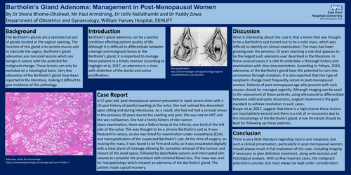
Bartholin’s Gland Adenoma: Management in Post-Menopausal Women By Dr Shona Bhome-Dhaliwal, Mr Paul Armstrong, Dr Jothi Nallathambi and Dr Paddy Zowa Department of Obstetrics and Gynaecology, William Harvey Hospital, EKHUFT
Background :The Bartholin's glands are a symmetrical pair of glands located at the vaginal opening. The function of this gland is to secrete mucus and to lubricate the vagina. Bartholin’s gland adenomas are rare solid lesions which are benign in nature with the potential for malignant change. These lesions can only be excluded on a histological basis. Very few adenomas of the Bartholin’s gland have been reported in the literature, making it difficult to give incidence of this pathology.
Introduction: Bartholin’s gland adenoma can be a painful condition affecting patient quality of life. Although it is difficult to differentiate between a benign and malignant lesion at the Bartholin's gland; it is important to manage these patients in a timely manner. According to Haghighi et al, 2017, an adenoma is a mass with distortion of the ductal and acinar proliferation.
Case Report: A 57-year-old, post-menopausal woman presented to rapid access clinic with a 10-year history of painful swelling at the vulva. She had noticed the discomfort upon sitting and during intercourse. As a result, she had not had a cervical smear in the previous 10 years due to the swelling and pain. She was not on HRT and she was nulliparous. She had a family history of skin cancer. Upon examination, there was a 9x6cm lump at the inferior, one third of the left side of the vulva. This was thought to be a chronic Bartholin's cyst as it was fluctuant in nature, so she was listed for examination under anaesthesia (EUA) and marsupialisation of the suspected Bartholin’s cyst. At the time of surgery, on incising the mass, it was found to be firm and solid, so it was enucleated digitally with a clear plane of cleavage allowing for complete removal of the tumour and closure of the dead space, followed by absorbable sutures and interrupted skin sutures to complete the procedure with minimal blood loss. The mass was sent for histopathology which showed an adenoma of the Bartholin’s gland. The patient made a good recovery.
Discussion: What is interesting about this case is that a lesion that was thought to be a Bartholin’s cyst turned out to be a solid mass, which was difficult to identify on clinical examination. The mass had been growing over the previous 10 years reaching a size that appears to be the largest such adenoma ever described in the literature. In these unusual cases it is vital to undertake a thorough history and examination with clear documentation. According to Yahaya, 2020, adenomas of the Bartholin’s gland have the potential to turn into carcinomas through mutation. It is also reported that this type of neoplastic change most frequently occurs in post-menopausal women. Patients of post-menopausal age who present with such masses should be managed urgently. Although imaging can be used in the assessment of these patients, using ultrasound to differentiate between solid and cystic structures, surgical treatment is the gold standard to achieve resolution in such cases. Burger et al, 2017, suggest that there is a high chance these lesions are incompletely excised and there is a risk of re-occurance due to the morphology of the Bartholin’s gland. A low threshold should be kept for following up these patients.
Conclusion :There is very little literature regarding such a rare neoplasia, but such a clinical presentation, particularly in post-menopausal women, should always result in full evaluation of the case, including imaging if necessary to plan definitive treatment, along with excision and histological analysis. With so few reported cases, the malignant potential is unclear, but must always be kept under consideration.