1,830 posters, 67 videos, 75 audios, 29 topics, 22 sessions, 4,727 authors, 1,697 institutions
ePostersLive by SciGen Technologies S.A. All rights reserved.
RCOG World Congress 2023
June 12 - 14, 2023 | London, UK

EP.0749
SUCCESFULL OUTCOME FOLLOWING DIAGNOSIS OF SUCCENTURIATE PLACENTA,VELAMENTOUS CORD INSERTION,VASA PRAEVIA.
Poster Presenter
Part of Topic
High Risk Pregnancy
SUCCESSFUL OUTCOME FOLLOWING DIAGNOSIS OF SUCCENTURIATE PLACENTA, VELAMENTOUS INSERTION OF THE UMBILICAL CORD AND VASA PRAEVIA : A CASE REPORT
Pinnaduwa S 1 , Ketheswarampillai1 , Marleen S1
1Sri Jayewardenepura General Hospital- Sri Lanka
Introduction
Morphological abnormalities of the placenta are rare but can result in increased maternal and fetal mortality and morbidity.1 Succenturiate placenta is the presence of one or more small accessory lobes that develop in the membranes at a distance from the periphery of the main placenta, to which they usually have vascular connections of fetal origin.1,2 Maternal age >35 years and pregnancies resulting from in vitro fertilization increase the risk of a succenturiate placenta. Velamentous cord insertion (VCI) is an abnormal cord insertion in which the umbilical vessels diverge as they traverse between the amnion and chorion before reaching the placenta. VCI is strongly associated with vasa praevia (VP), where umbilical vessels lie in close proximity to the internal cervical os. VP leaves the vessels vulnerable to rupture, which can lead to fatal fetal exsanguination. Only 1.6% of succenturiate placentas are associated with VCI and VP and carry fetal mortality ranging from 33% to 100%.3,4,5 This case report is a successful antenatal ultrasound detection of a succenturiate placenta with VCI and VP and timely planned delivery minimizing potential complications leading to a successful pregnancy outcome.
Case
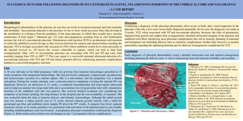
A 36 year old lady in her third pregnancy with two previous first trimester miscarriages presented at 31 weeks gestation with antepartum haemorrhage. She had previously undergone a laparoscopic myomectomy and hysteroscopic resection of a uterine septum. She is a non-smoker, and her pregnancy was a natural conception. During her routine anomaly scan, a placenta praevia completely covering the internal OS was diagnosed. However, on admission at 31 weeks, a combined transabdominal and transvaginal ultrasound scan revealed an anterior low-lying main lobe and a succenturiate low-lying posterior lobe with velamentous insertion of the umbilical cord and vasa praevia. She received tailored in-patient care considering her symptoms of persistent spotting, the long distance to the hospital and the non-availability of transport. She was counseled about the risks of preterm delivery and obstetric haemorrhage. Although the initial growth scan was normal, a repeat growth scan at 33 weeks showed reduced growth velocity with a small for gestational age fetus and umbilical artery doppler PI above the 95 th centile. A category four lower segment caesarean section at 34 weeks gestation was performed with activation of the placenta praevia care bundle, including multidisciplinary team involvement. A postpartum placental examination confirmed the diagnosis (Figure 1). Both mother and newborn were discharged on day 12 postpartum with no complications.
Discussion
Confirming a diagnosis of this placental abnormality often occurs at birth, after visual inspection of the placenta, with only a few cases successfully diagnosed antenatally. In our case, the diagnosis was made at 31weeks. VCI, when associated with VP and succenturate placenta, increases the risks of prematurity, impaired fetal growth and sudden fetal exsanguination. Detailed ultrasound imaging of the placenta and umbilical cord allow identifying most placental complications thus aid in strategic planning of antenatal and intrapatum care including delivery time to minimise complications. Further data, however, is needed, particularly regarding the optimal gestational age for delivery of pregnancies complicated by VCI.
Conclusion
Early diagnosis of placental abnormalities using a detailed ultrasound scan and optimal management, including planning the delivery time, is vital in minimizing fetal and maternal morbidity and mortality.
References
1. Obstetrics, W., 2005. Cunningham EG, Leveno KJ, Bloom SL, Hauth JC, Gilstrap L, Wenstrom KD, editors 22nd ed.
2. Suzuki, S. and Igarashi, M., 2008. Clinical significance of pregnancies with succenturiate lobes of placenta. Archives of gynecology and obstetrics, 277(4), pp.299-301.
3. Stelzl, P.W., Yadav, G., Perley, L. and Silasi, M., 2017. Early sonographic detection of a succenturiate placenta after IVF in a 42-year-old woman with multiple comorbidities. Case Reports, 2017, pp.bcr-2017
4. Moreno Santillán, A.A., Arriaga López, A. and Martínez Adame, L.M., 2018. Placenta succenturiata. Reporte de un caso. Revista de la Facultad de Medicina (México), 61(2), pp.37-41.
5. Shukunami, K.I., Tsunezawa, W., Hosokawa, K., Tajima, K. and Kotsuji, F., 2001. Placenta previa of a succenturiate lobe: a report of two cases. European Journal of Obstetrics & Gynecology and Reproductive Biology, 99(2), pp.276-277