1,830 posters, 67 videos, 75 audios, 29 topics, 22 sessions, 4,727 authors, 1,697 institutions
ePostersLive by SciGen Technologies S.A. All rights reserved.
RCOG World Congress 2023
June 12 - 14, 2023 | London, UK

EP.0734
Pyomyoma complicated by utero-cutaneous fistula - successful uterine conservation by secondary intention healing
Poster Presenter
Authors
Ming Fen Grace Chan, Soe-na Choo, Shuang Ye Pearl Tong, Lin Zhenjia Harvard
Affiliations
Part of Topic
Benign Gynaecology
Pyomyoma complicated by utero-cutaneous fistula - successful uterine conservation by secondary intention healing
Grace MF Chan1, Soe-na Choo1, Pearl SY Tong1, Harvard Lin1
1 Department of Obstetrics and Gynaecology, National University Hospital, 1E Kent Ridge Road, Singapore 119228, Singapore
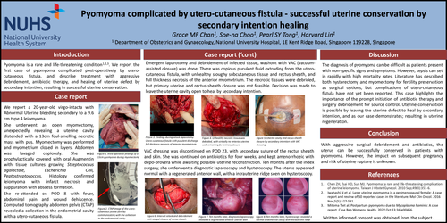
Pyomyoma is a rare and life-threatening condition1,2,3. We report the first case of pyomyoma complicated post-operatively by utero-cutaneous fistula, and describe treatment with aggressive debridement, antibiotic therapy, and healing of uterine defect by secondary intention, resulting in successful uterine conservation.
We report a 20-year-old virgo-intacta with Abnormal Uterine bleeding secondary to a 9.6 cm type 4 leiomyoma. She underwent an open myomectomy, unexpectedly revealing a uterine cavity distended with a 13cm foul-smelling necrotic mass with pus. Myomectomy was performed and myometrium closed in layers. Abdomen was lavaged copiously. She was prophylactically covered with oral Augmentin with tissue cultures growing Streptococcus agalactaie, Escherichia Coli, Peptostreptococcus. Histology confirmed leiomyoma with infarct necrosis and suppuration with abscess formation. She re-attended on POD 8 with fever, abdominal pain and wound dehiscence. Computed tomography abdomen pelvis (CTAP) revealed a collection in the endometrial cavity with a utero-cutaneous fistula.
Emergent laparotomy and debridement of infected tissue, washout with VAC (vacuum-assisted closure) was done. There was copious purulent fluid extruding from the utero-cutaneous fistula, with unhealthy sloughy subcutaneous tissue and rectus sheath, and full thickness necrosis of the anterior myometrium. The necrotic tissues were debrided, but primary uterine and rectus sheath closure was not feasible. Decision was made to leave the uterine cavity open to heal by secondary intention.
VAC dressing was discontinued on POD 23, with secondary suture of the rectus sheath and skin. She was continued on antibiotics for four weeks, and kept amenorrhoeic with depo-provera while awaiting possible uterine reconstruction. Ten months after the index surgery, she underwent a diagnostic laparoscopy and hysteroscopy. The uterus appeared normal with a regenerated anterior wall, with a intrauterine ridge seen on hysteroscopy.
The diagnosis of pyomyoma can be difficult as patients present with non-specific signs and symptoms. However, sepsis can set in rapidly with high mortality rates. Literature has described both hysterectomy and myomectomy for fertility preservation as surgical options, but complications of utero-cutaneous fistula have not yet been reported. This case highlights the importance of the prompt initiation of antibiotic therapy and surgery debridement for source control. Uterine conservation is possible by leaving the uterine defect to heal by secondary intention, and as our case demonstrates; resulting in uterine regeneration. With aggressive surgical debridement and antibiotics, the uterus can be successfully conserved in patients with pyomyoma. However, the impact on subsequent pregnancy and risk of uterine rupture is unknown.