1,830 posters, 67 videos, 75 audios, 29 topics, 22 sessions, 4,727 authors, 1,697 institutions
ePostersLive by SciGen Technologies S.A. All rights reserved.
RCOG World Congress 2023
June 12 - 14, 2023 | London, UK

EP.0728
A Case of Granulomatous Mastitis and Erythema Nodosum (GMEN) in Pregnancy
Poster Presenter
Authors
Meei Jiun Seet, E-Jyn Michelle Kwek, Jia Ling Cheryl Lie, Swee Ho Lim
Affiliations
Part of Topic
Maternal Medicine
A Case of Granulomatous Mastitis and Erythema Nodosum (GMEN) in Pregnancy
Granulomatous mastitis (GM) is a rare benign, inflammatory condition of the breast, more commonly found in parous women who had given birth in the last five years (1). It has a prevalence of 2.4 per 100000 women aged 20-40 years with a mean age of 34 years (2). Erythema nodosum (EN) is an inflammatory disorder affecting the subcutaneous fat, characterised by tender, erythematous nodules, and can be a rare extramammary manifestation of GM.
Although the pathogenesis for both GM and EN is unknown, possible etiologies include pregnancy, infection, sarcoidosis and autoimmune disease. Here, we report a case of granulomatous mastitis with erythema nodosum (GMEN) in pregnancy, possibly related to latent tuberculosis (TB).
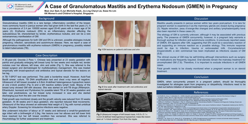
A 36-year-old, Gravida 2 Para 1, Chinese lady presented at 23 weeks gestation with painful and gradually enlarging left breast lump for two weeks and multiple red, tender nodules over her elbows, left knee, shin and ankle (Fig 1). She was referred to the breast surgeon and dermatologist for multidisciplinary management. She was treated empirically with broad-spectrum oral antibiotics and topical steroids for the lesions on her leg.
A TB T-SPOT test was performed. This yield a borderline result. However, Acid-Fast Bacilli smear, culture, TB DNA amplification test and chest x-ray were all negative. Ultrasound of left breast revealed a 6.8cm ill-defined heterogeneous hypoechoic lesion at one o’clock position (Fig 3) and a left reactive axillary lymph node. Biopsy of the breast lump showed GM with abscess. She was started on anti-TB drugs (Rifampicin, Ethambutol, Isoniazid and Pyridoxine) for possible latent TB at 30 weeks gestation and later, oral prednisolone, as her breast lump increased in size and she started discharging pus from the skin over the breast.
Fetal growth was monitored closely and the growth velocity was reduced from 33 weeks gestation. At 36 weeks and 5 days gestation, she reported reduced fetalmovements. Ultrasound of the fetusshowed an estimated fetal weight of 2.1kg with normal umbilical artery Doppler. Screening for intrauterine infections were negative.
She underwent induction of labour at 37 weeks and delivered a small-for-gestational-age infant with a birthweight of 2.2kg. Her lesions over her elbows and left lower limb have resolved but her left breast condition has worsened. She was referred to rheumatology for further assessment and treatment.
Mastitis usually presents in parous women within two years post-partum. It is rare for pregnant women to experience it as the mammary glands are closed during pregnancy (3). Nipple retraction, peau d’orange-like changes and axillary lymphadenopathy have also been reported in these cases (4).
The etiology of GM is currently unknown, although it may be associated with previous parity. The presence of GMEN concurrently, however, in a pregnant lady warrants a thorough workup for infection and autoimmune conditions. In previously reported cases of GMEN, EN appears after GM, suggesting that EN could be a secondary reaction of and supporting an immune reaction as a possible etiology. This immune response could be due to infection, trauma or extravasated milk. Corynebacterium kroppenstedtii, a skin commensal has been implicated as infections causes of GM (5,6).
The clinical course of GM may be self-limiting although interventions such as surgery or medications are frequently required. Oral steroids remain the mainstay treatment for uncomplicated GM (7,8). Therefore, it is important to exclude infections in all GMEN cases.
Fetal growth needs to be closely monitored when a pregnant woman is on steroid treatment.
GMEN, when concurrently present in a pregnant patient, should be thoroughly investigated for etiologies apart from pregnancy or idiopathicity. Infections need to be ruled out before initiation of steroid treatment.