1,830 posters, 67 videos, 75 audios, 29 topics, 22 sessions, 4,727 authors, 1,697 institutions
ePostersLive by SciGen Technologies S.A. All rights reserved.
RCOG World Congress 2023
June 12 - 14, 2023 | London, UK

EP.0709
How predictable? Presenting as an Emergency!
Poster Presenter
Authors
Affiliations
Part of Topic
Gynae Oncology
Introduction
•Vulval intraepithelial neoplasia (VIN) is considered a pre-malignant condition with risk factors including human papilloma virus (HPV), smoking, and lichen sclerosis.
•
•It is a histological diagnosis hence a biopsy is required and this case clearly demonstrates the importance of prompt biopsy especially as there was uncertainty in the clinical diagnosis.
•
•Classification of stages of VIN progress from VIN 1 to VIN 3 and the focus in this case was that of VIN 2.
Case Report
•43-year-old woman presented to the accident and emergency (A&E) department with an 18-month history of progressively increasing vulva pain, itching, and swelling. There was also a history of recent unintentional weight loss and smoking. She had trialled emollients and antifungal creams with no relief of symptoms. She had a previous history of cone biopsy and her last smear test a year prior to presentation was positive for HPV.
•Examination showed tender, bilaterally inflamed, and protruding inferior labial minora and majora. There were also areas of leucoplakia on the posterior fourchette. Inflammatory markers were normal.
•Biopsy was taken by the Gynaecology team in A&E and steroids commenced for possible chronic inflammatory conditions such as lichen sclerosis.
•Histology result showed VIN 2 with positive expression for P16. She was then reviewed by a Gynaecologist with a special interest in Gynae-oncology following which a wide local excision (WLE) of the vulva was done. Histology following WLE confirmed an extensive complex high-grade VIN 2 and 3 with P16 block positivity in keeping with HPV-associated disease. There was no evidence of invasive malignancy.
Discussion
•VIN is increasingly becoming more reported amongst women in their forties. A high index of suspicion is therefore needed when faced with confusing clinical scenarios.
•An assessment of risk factors and correlation with current problems might lead to a diagnosis. A simple and effective way to diagnose early is by taking a biopsy of concerning lesions without delay.
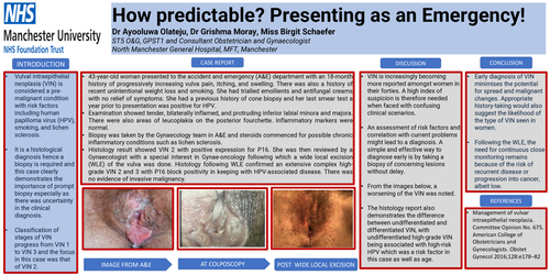
•From the images below, a worsening of the VIN was noted.
•The histology report also demonstrates the difference between undifferentiated and differentiated VIN, with undifferentiated high-grade VIN being associated with high-risk HPV which was a risk factor in this case as well as age.
Conclusion
•Early diagnosis of VIN minimises the potential for spread and malignant changes. Appropriate history-taking would also suggest the likelihood of the type of VIN seen in women.
•
•Following the WLE, the need for continuous close monitoring remains because of the risk of recurrent disease or progression into cancer, albeit low.
References
•Management of vulvar intraepithelial neoplasia. Committee Opinion No. 675. American College of Obstetricians and Gynecologists. Obstet Gynecol 2016;128:e178–82