1,830 posters, 67 videos, 75 audios, 29 topics, 22 sessions, 4,727 authors, 1,697 institutions
ePostersLive by SciGen Technologies S.A. All rights reserved.
RCOG World Congress 2023
June 12 - 14, 2023 | London, UK

EP.0682
Peritoneal Melanosis with Bilateral Serous Cystadenomas of the Ovaries: A Case Report
Poster Presenter
Part of Topic
Benign Gynaecology
Peritoneal Melanosis with bilateral serous cystadenomas of the ovaries: A Case Report
Objective
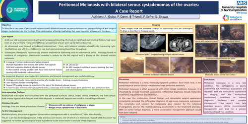
To describe a rare case of peritoneal melanosis with bilateral ovarian serous cystadenomas, using radiological and surgical images to demonstrate the findings This combination of benign pathology has been reported only once in literature.
Case Report
A 60 year old woman presented with postmenopausal bleeding She had no significant past medical history, had never been on any hormone replacement therapy and cervical smears were up to date and normal.
An ultrasound scan showed a thickened endometrium-7 mm, with bilateral complex adnexal cysts, measuring right 24 x 20 x 26 mm and left 51 x 41 x 46 mm in size, both demonstrating blood flow throughout. Subsequent therapeutic hysteroscopy showed endometrial thickening and an endometrial polyp. Histology found no evidence of malignancy. Examination revealed a nodule on the left vaginal wall- a biopsy of this showed nodular melanosis.
A staging CT ( abdomen and pelvis) showed
-Multiple hypodense liver lesions with a thick liver capsule
-Solid and suspicious bilateral adnexal masses
-A thick walled urinary bladder with focal intra luminal lesions causing mild-moderate hydronephrosis
-CA 125 was 27
-An MRI revealed multifocal lesions involving the liver and adrenal glands
The suspected diagnosis was metastatic melanoma and onward management was multidisciplinary:
-Urology performed a transurethral resection of a bladder lesion- histology revealed melanosis
-Liver biopsies showed no malignancy
-Repeat CT imaging showed a reduction in the size of the bilateral adnexal masses
-A laparoscopic bilateral salpingo-oophorectomy, cystoscopy and bladder biopsy were performed as a joint procedure.
Intra-operative findings:
Widespread black spots were visualised over the peritoneal surfaces, uterus, bowel serosa, omentum, and liver surface. Both ovaries appeared multicystic with black deposits A melanotic deposit was noted in the left vaginal fornix.
Histology Results:
Histology from the above procedures showed melanosis with no evidence of malignancy or atypia and benign serous cystadenomas of the ovaries.
Further Management:
Multidisciplinary discussion suggested conservative management with a surveillance CT scan in six months. This CT scan has showed progression in the previous seen lesion, one of which is in the breast. Repeat MDT discussion has suggested no further gynaecological input but referral to the breast team to exclude other diagnoses.
Discussion
Peritoneal melanosis is a rare, minimally reported condition. Even more rare, is the combination of ovarian serous cystadenomas with peritoneal melanosis.
Peritoneal melanosis is often associated with other benign conditions however, it is important to exclude malignant associations. Differential diagnoses include metastatic melanoma and peritoneal endometriosis. In this case, the multisystem clinical findings and remarkable surgical appearance immediately provoked the differential diagnosis of aggressive melanoma metastases. The complexity and concern for malignancy gave reasons for the extensive investigations and for the multidisciplinary involvement in the treatment provided. With an earlier benign diagnosis, a more conservative management approach would have been adopted.
Conclusion
Peritoneal melanosis is a very rare condition. The pathogenesis is poorly understood but numerous associations are reported. Both the non specific appearance on imaging and the multisystem involvement, contribute to indirect diagnostic pathways and complex management. Case reports may help postulate causes, define recommended management and lead to a greater understanding of this condition.
References
1.Mhand M, Obed R, Harouachi A, Haloui A, Bouhout T, Bennani A, Serji B, Harroudi TE. Peritoneal melanosis associated with serous carcinoma of the ovary: A case report and literature review. Ann Med Surg Lond ). 2022 Aug 18;81:104452. doi : 10.1016/j.amsu.2022.104452. PMID: 36147165; PMCID: PMC9486698.
2. Kim NR, Suh YL, Song SY, Ahn G. Peritoneal melanosis combined with serous cystadenoma of the ovary: a case report and literature review. Pathol Int. 2002 Nov;52(11):724 9. doi : 10.1046/j.1440 1827.2002.01405.x. PMID: 12685549.