1,830 posters, 67 videos, 75 audios, 29 topics, 22 sessions, 4,727 authors, 1,697 institutions
ePostersLive by SciGen Technologies S.A. All rights reserved.
RCOG World Congress 2023
June 12 - 14, 2023 | London, UK

EP.0578
Status Epilepticus associated to Ovarian Teratoma
Poster Presenter
Authors
Affiliations
Part of Topic
Benign Gynaecology
Anti-NMDAR encephalitis associated with ovarian teratoma: A case report
Nourah Al Kindi*, Shabana Kapadia, Wafa Talha, Rana Al Alami
Obstetrics and Gynecology department, Tawam Hospital, United Arab Emirates
Introduction
Anti-N-Methyl-D-Aspartate Receptor (NMDAR) encephalitis is a subcategory of auto-immune encephalitis. It can be secreted by ovarian teratoma, which cause serious and fatal pathology in young women. Unfortunately, this association is underrecognized by gynecologists as patients usually presenting with psychiatric and neurological symptoms. There were total of 174 cases of ovarian teratoma-associated anti-NMDAR encephalitis reported worldwide, but current case considers the first from United Arab Emirates. We reported it to increase awareness over this pathology and highlight on the challenges of the diagnosis and treatment of it.
Case report
A 30-year-old Ugandan house-maid Para 1, full term spontaneous vaginal delivery with no significant past medical or surgical history. Brought to Emergency Department with abnormal behavior, history of aggressiveness, labile mood with irrational talk for one day. Initial investigations excluded any organic causes of her abnormal behavior. Computerized Tomography (CT) of the brain was normal. Admitted under psychiatry with impression of acute psychosis for further treatment and management.
During admission:
She had two episodes of a generalized tonic clonic seizure associated with urinary incontinence and frothy discharge from her mouth. Repeated investigation showed increased inflammatory markers, suspected central nervous system (CNS) infection. Neurological examination and Magnetic Resonance imaging (MRI) of the brain were normal.
In view of that, patient was started on antiepileptic medications and transferred to stroke unit for an electroencephalogram (EEG) which reported encephalopathic record. Then, lumbar puncture proceeded which reported mild lymphocytic pleocytosis with normal protein and glucose with impression of infectious vs autoimmune encephalitis. Steroid, intravenous immunoglobulin and antiviral medications given and autoimmune screen requested.
On day five of the admission:
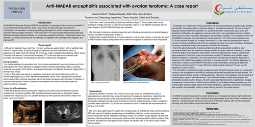
Patient developed recurrent seizures (status epilepticus) with altered consciousness which required intubation with mechanical ventilation. MRI and ultrasound pelvis performed and reported left ovarian complex mass measuring 3.5x2.4 cm, possible dermoid cyst with significant amount of fluid noted in the pelvis. Figure 1 and figure 2.
Furthermore, chest X-ray showed right side pleural effusions (figure 3). Tumor markers were normal. Impression of Meigs syndrome vs autoimmune encephalitis -features of anti NMDAR encephalitis were on board. Based on that Anti NMDAR antibody was requested.
After that, patient underwent exploratory laparotomy with left salpingo-ophorectomy and omental biopsy as she was unsuitable for laparoscopy (Figure 4).
Histopathology revealed single focus of immature element in mature cystic teratoma. In few days, the report of NMDA receptor antibody came positive which confirms the diagnosis of autoimmune limbic encephalitis
Postoperatively:
patient was admitted to Intensive Care Unit (ICU) for 42 days where she underwent five cycles of plasmapheresis on alternative days along with adjustment of antiepileptic medications. Despite that, the patient had recurrent seizures which required tracheostomy, two doses of rituximab, adjustment of antiepileptic medications another course of steroids and monthly cyclophosphamide. Further investigations confirmed latent tuberculosis (TB), so she was commenced on anti-TB treatment with recommendation to continue for 9 months.
After seven days, patient was off sedation with no seizures and was shifted to the stroke unit where she had PEG tube insertion for feeding, physiotherapy and rehabilitation. After four months, tracheostomy was removed and patient started mobilization, feeding by herself, but sometimes she got agitated with abnormal behaviour. Neuropsychological screening was performed which reported persistent cognitive problems. After 6 months, the patient was discharged from the hospital, she travelled back to her home country in stable clinical condition.
Discussion
Encephalitis is an inflammation of brain parenchyma with neurologic symptoms resulting in major morbidity and mortality. It arises from either an infectious etiology or an autoimmune disorder. The anti-N-methyl-D- aspartate receptor (NMDAR) encephalitis is an autoimmune disorder characterized abrupt onset, with psychiatric manifestations like aggression, psychosis and memory loss, associated to seizures, cognitive decline and coma which may require tracheostomy and mechanical ventilation. 57% of published cases with anti-NMDAR encephalitis were associated with mature teratomas, 16.7% with immature teratomas and 3.4% with immature teratoma foci. However, there were 23% of the cases the histological type was not reported. Teratomas exhibit elements of the three blastodermic layers and contain neural tissue that triggers an immune response resulting in over-production of anti-NMDAR antibodies.
Most of the patients have same clinical presentation including neuropsychiatric symptoms, rapid neurological deterioration and behavioural changes included psychosis, abnormal movements of the face, mouth and tongue, fever, headache, hallucinations, rhinorrhea, hypersalivation, seizures, catatonia and status epilepticus. Furthermore, most of reported cases required ICU admission and ventilator support before the diagnosis is established. The clinical presentation may mis-lead and delay the diagnosis and treatment of anti- NMDAR encephalitis to psychiatric or neurologic disorder. The definitive diagnosis is by confirmation of presence of anti-NMDAR antibodies in blood or preferably in cerebrospinal fluid (CSF). Furthermore, abdomino-pelvic CT image, MRI pelvis and transvaginal ultrasound (TVU) are used for diagnosis of ovarian teratoma.
The combination of medical and surgical management yields the best therapeutic results and a more rapid recovery in the management of anti-NMDAR antibodies encephalitis associated with ovarian teratoma. The medical treatments include acyclovir, methylprednisolone, intravenous immunoglobulin, anticonvulsants and chemotherapy in cases of immature teratoma. Other effective therapies included plasmapheresis and antiepileptic and anticonvulsive therapy, with some cases requiring tracheostomy or gastrostomy. Surgical management include removal of mature teratoma with/without uni or bilateral salpingo-oophorectomy, and radical surgery was reserved for patients with immature teratomas. Additionally, recurrence of the disease was reported in few cases after months to years following removal of the ovarian teratoma.
Conclusion
Anti-NMDA encephalitis associated to ovarian teratoma is a rare disease that carries a high mortality and morbidity in young women. It is under-recognized among gynecologist which lead to delay in diagnosis and treatment. Literature reported full recovery was noticed in 80% of the patients, after 3 months of hospital discharge, who had combined surgical and medical treatment.