1,830 posters, 67 videos, 75 audios, 29 topics, 22 sessions, 4,727 authors, 1,697 institutions
ePostersLive by SciGen Technologies S.A. All rights reserved.
RCOG World Congress 2023
June 12 - 14, 2023 | London, UK

EP.0573
VULVAR SCHISTOSOMIASIS; AN IMPORTANT BUT UNCOMMON DIAGNOSIS, “A NEAR MISS”
Part of Topic
Benign Gynaecology
Vulvar Schistosomiasis: An Important but Uncommon Diagnosis: “A Near Miss”.
Eze I.O 1 , Ugbem T.I3, Archibong E.I2 , Elegbua C.O2 , Afolayan S.T2 , Meremikwu C.A 2
Introduction
Vulvar schistosomiasis, part of the spectrum of female genital schistosomiasis (FGS), is an important tropical disease with far-reaching negative impact on female reproductive health. However, this disease is rarely diagnosed and reported in clinical setting even in endemic tropical Africa like Nigeria due to poor awareness and low index of suspicion among the physicians in the tropics. This could result in avoidable morbidities
CASE SUMMARY
A 23-year-old graduate who presented with 6 months history of vulvar itching and swelling. Itching was intermittent but intense, causing her significant discomfort, a times leading to bruising and occasional embarrassment. There was no known aggravating factor. Vulva swelling located around the labia minora was noticed about the same period. It felt rubbery, occasionally rough but was not painful. There was no associated abnormal vaginal discharge or odour. No urinary symptoms was reported by patient. No history of travel to a high endemic schistosomiasis zone.
She had indulged in self-medication with a lot of topical creams and vaginal inserts to no avail. She Presented to the gynaecology clinic of a reference hospital in southern Nigeria for assessment.
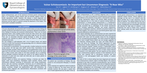
On examination at presentation, she was generally a healthy looking but worried young woman. All vital signs were within normal limits. Examination of the external genitalia revealed a fleshy, partly warty and papillomatous lesion on the upper lip of the left labia minora extending to the left lower base of the clitoris (Fig 1). Minor bruises were noted on the lesion (possibly due to scratching). Mucoid (probably ovulatory) discharge was noted on the vaginal introitus. An impression of vulval wart was made and initial plan was to place patient on topical podophyllin cream.
However, based on history and suspicious findings, a decision was taken for biopsy under local anaesthesia. Histological report revealed “intense mixed inflammatory infiltrates consisting of mononuclear cells mainly lymphocytes, plasma cells and numerous eosinophils. Numerous ova of schistosomes also noted within collections.(Fig. 2) The overlying stratified squamous epithelium displays acanthotic changes and papillomatosis with mild koilocytosis. No dysplasia noted. Diagnosis was “granulomatous inflammation consistent with Schistosoma granuloma
MANAGEMENT/INTERVENTION
The patient was counselled on findings. She was started on oral praziquantel 40mg/kg in 3 divided doses. Patient reported very significant relief of symptoms 2 weeks on follow up. The vulvar lesion also had markedly resolved. She was given a repeat dose and a 4-week appointment but she didn’t show up
DISCUSSION
Vulvar schistosomiasis is not a common diagnosis even in endemic regions.1, 2 It is part of the spectrum of female genital schistosomiasis (FGS) defined as the presence of ova and/or a characteristic pathology in female reproductive organs.4 FGS remains highly prevalent and under-diagnosed due to a low index of suspicion among health-care professionals.5, 6 Female genital schistosomiasis is a manifestation mainly of the species S. haematobium infection. Several studies have strongly associated FGS to pathologies of the female reproductive system including subfertility, infertility, and ectopic pregnancies, pregnancy complications (abortion, still-birth and preterm delivery and low birth weight).1 The uterine cervix is the female genital organ most frequently affected by schistosomiasis.3, 6
Vulvo-vaginal pathology due to schistosomiasis has been reported for over a century 3 Our index patient presented with vulvar lesion grossly consistent with vulvar wart the possible reason for the misdiagnosis even by unsuspecting gynaecologists. We nearly missed the diagnosis but for more in-depth history (which was not strongly suggestive of HPV infection), thinking outside the usual, and timely decision to biopsy the lesion for histology. The clinical appearance of vulval schistosomiasis includes progressive and relapsing swelling, painful or painless ulceration, papules, nodules, pruritus, a hypertrophic clitoris with an eroded granular surface and papillomatous lesions forming masses resembling condylomata.5,6 Some of these clinical features were present in our index patient. The vast majority are from women living in schistosomiasis endemic countries or from individual cases of tourists who acquired FGS in sub-Saharan countries and were diagnosed in the well-equipped health care facilities of western countries.7
Few cases of FGS in clinical setting have been diagnosed and reported from schistosomiasis endemic regions including Nigeria. This could be attributed to low index of suspicion among physicians, lack of reliable diagnostic facilities and trained personnel, lack of integrated approach to diagnosis and treatment. Hence, a lot of such cases may have been neglected, misdiagnosed and mismanaged as other common genital pathologies that physicians are used to. This could result to significant but avoidable morbidities and mortalities.
CONCLUSION
Female genital schistosomiasis is an important but neglected tropical disease. Clinical diagnosis and reportage are few even in an endemic zone like Nigeria possibly due to poor awareness and low index of suspicion. The authors believe this case is one of numerous cases of FGS that may have been missed, misdiagnosed and mismanaged, and hence calls attention for high index of suspicion. It also emphasizes the need to biopsy every suspicious lesion without which this case would have been missed.