1,830 posters, 67 videos, 75 audios, 29 topics, 22 sessions, 4,727 authors, 1,697 institutions
ePostersLive by SciGen Technologies S.A. All rights reserved.
RCOG World Congress 2023
June 12 - 14, 2023 | London, UK

EP.0499
X-linked hypophosphatemia in pregnancy. Very rare Case series with management outline.
Poster Presenter
Authors
Affiliations
Part of Topic
Maternal Medicine
X-linked Hypophosphatemia in Pregnancy: Case Series with Management Outline
Dr M Christodoulou, Mr A Aref, Dr H Mahmoud
Introduction
Familial X-linked hypophosphataemia rickets1
-
Rare inherited condition of bone metabolism
-
Affects 1 in 20,000 individuals
-
Caused by a mutation in the PHEX gene resulting in
-
Excess production of FGF23 and thus (2)
-
Excessive urinary excretion of phosphate
-
-
50% risk of transmission to offspring
Bone Metabolism
Interactions between PTH, calcitonin and VitD as depicted in image 1
Role of FGF23 in calcium homeostasis was only recently discovered
➢ its function is to prevent VitD mediated hyperphosphatemia
➢ Net effect of FGF23 is to decrease phosphate2
Pregnancy
-
Is a period of increased calcium requirement to encourage growth of foetal tissues
-
Most of calcium and phosphate transport occurs in the third trimester against a concentration gradient2.
Aim:
Case series of two sisters with XLH followed up during pregnancy at Medway Hospital from October 2021 to December 2022 with an aim to highlight the management and outcome of this rare condition in pregnancy.
Design and Methodology
Timescale: October 2021 to December 2022
Methods: Electronic records of their pregnancies were reviewed, including antenatal clinics, emergency presentations and admissions as well as delivery outcomes.
Analysis: Excel was used for calculations. Results and management compared to PubMed literature using search words.
Results
Booking phosphate levels 0.34mmol/L and 0.4mmol/L Antenatal planning
Medication: Alfacacidol, PO Phosphate & Aspirin 150mg due to high risk of IUGR.
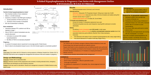
Serum phosphate levels measured every 3wks in first two trimesters and weekly during third trimester (trends shown in graph 1)
Additional fetal growth scans on weeks 28, 32 and 36wks 3rd trimester and intrapartum care
During 3rd trimester IV phosphate replacement was used.
Parenteral phosphate was also used intrapartum requiring MCU admission to monitor electrolytes.
Pre-delivery levels of phosphate were 0.52 mmol/L in both cases
Planned delivery method at 37wks by elective uncomplicated caesarean section due to the associated rickets bony pelvis affection (Rachitic Pelvic Osteo-dystrophy).
First delivered at 36 weeks+3 due to resistant hypophosphatemia with a baby weight 2920 g.
The second delivered at 37 weeks+2 with baby weight 2725 g.
Protocol for IV phosphate replacement
-
Admission to MECU and overnight stay
-
IV infusion of Monofer iron
-
Followed by an infusion pump of 30mmol of
phosphate infusion over 12hrs with a maximum infusion rate of 10 mmol/hr to avoid hypocalcaemia.
-
Four hourly VBGs were performed to monitor calcium levels.
-
Overnight stay and repeat bloods next morning with aim of phosphate > 0.3 mmol/L
In the case of resistant hypophosphataemia, the reinfusion included IV phosphate 50mmol over 12hrs using infusion pump at rate of 10 mmol/hr.
Conclusions
-
Two relatives with XLH-Rickets managed at Medway Maternal-Medicine-Centre with Endocrinology Input with good neonatal and maternal outcome.
-
Regular blood tests for serum phosphate levels and additional growth scans performed
-
Aim to keep IV phosphate use limited for third trimester to reduce risk of hypocalcaemia
-
Increased demand of phosphorus in 3rd trimester is in keeping with literature4
-
Delivery via ELCS
-
Limited previous literature demonstrated similar management5
Limitations
-
No control of confounding factors
-
Small number of participants / difficult to generalise results
-
Young age of participants
-
No guidelines for requirement of IV phosphate; thus, clinical judgement had to be
used.
-
Limited literature to compare