1,830 posters, 67 videos, 75 audios, 29 topics, 22 sessions, 4,727 authors, 1,697 institutions
ePostersLive by SciGen Technologies S.A. All rights reserved.
RCOG World Congress 2023
June 12 - 14, 2023 | London, UK

EP.0424
MALIGNANT PHYLLODES TUMOR IN PREGNANCY: A CASE REPORT
Poster Presenter
Authors
Affiliations
Part of Topic
High Risk Pregnancy
MALIGNANT PHYLLODES TUMOR IN PREGNANCY: A CASE REPORT
INTRODUCTION
- Phyllodes tumor of the breast is a rare fibroepithelial neoplasm that accounts < 1% of all female breast neoplasms. Being a rare condition, the influence of pregnancy on the course of phyllodes tumor has not been documented.
- In the Philippines, there has been no reported case of malignant phyllodes tumor in pregnancy and there are no local literature available regarding this topic.
CASE REPORT
- A 32-year-old, female, G2P1 (1001) PU 32 weeks and 2 days AOG, not in labor, presented at the OPD department with a chief complaint of rapidly growing breast mass for the past 4-5 months.
- Past medical history, family history, personal and social history: unremarkable.
- First pregnancy was in 2016, delivered full term via NSD at home with no complications. Second pregnancy is the current pregnancy.
- Patient's menarche was at 12 years old, with an interval of 30 days, duration of 5 days, with use of 4 pads/day and with associated dysmenorrhea. She had her coitarche at 25 years old, with only one sexual partner, no associated dyspareunia, no history of STIs and with history of OCP use for 1 vear.
PHYSICAL EXAMINATION:
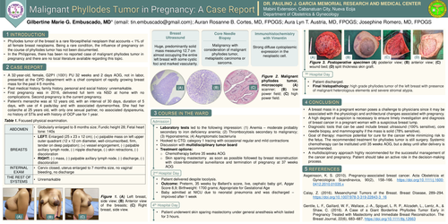
Abdomen: Globularly enlarged to 8 months size; Fundic height 28; Fetal heart tone: 140s
Breasts: Left Breast: enlarged to 25 × 23 × 12 cm with (+) palpable mass on left upper outer quadrant (12 × 12 cm diameter, well-circumscribed, firm, tender on deep palpation); (+) vessel engorgement, (-) palpable axillary lymph node, (-) nipple discharge, (-) skin retractions, (-) discoloration; Essentially normal right breast on physical examination
Internal Examination: Cervix closed, uterus enlarged to 7 months size, no vaginal bleeding, no discharge
DIAGNOSTICS:
Breast Ultrasound- Huge, predominantly solid mass measuring 12.7 cm almost occupying the entire left breast with some cystic foci and marked vascularity.
Core Needle Biopsy- Malignancy with consideration of malignant phyllodes tumor, metaplastic carcinoma or sarcoma.
Immunohistochemistry with vimentin- Strong diffuse cytoplasmic expression in the neoplastic cell.
COURSE IN THE WARD:
Upon admission, laboratory tests led to the following impression: (1) Anemia - moderate probably secondary to iron deficiency anemia; (2) Thrombocytosis secondary to malignancy;
(3) Hyponatremia; (4) Asymptomatic bacteriuria
- Hooked to CTG: category 1 tracing with occasional regular and mild contractions
Discussion with multidisciplinary tumor board - Treatment options of Chemotherapy before 35 weeks age of gestation vs. Skin sparing mastectomy as soon as possible followed by breast reconstruction with close-fetomaternal surveillance and termination of pregnancy at 37 weeks
AOG
On 2nd hospital day:
The patient delivered despite tocolysis.
Outcome: Preterm, 35 weeks by Ballard's score, live, cephalic baby girl, Apgar Score 8,9; Birthweight: 1700 grams; Appropriate for Gestational Age Baby admitted at NICU due to neonatal pneumonia and was discharged improved after 1 week
On 4th hospital day:
The patient underwent skin sparing mastectomy under general anesthesia which lasted for 3 hours.
On 5th hospital day:
The patient was discharged.
Final histopathology: high grade phyllodes tumor of the left breast with presence of malignant heterologous elements and severe stromal atypia
CONCLUSION:
- A breast mass in a pregnant woman poses a challenge to physicians since it may be associated with the physiologic and architectural changes associated with pregnancy.
- A high degree of suspicion is necessary to ensure timely investigation and diagnosis of breast cancer in a pregnant woman with a suspicious breast lump.
- Diagnostic tools that can be used include breast ultrasound (100% sensitive), core needle biopsy, and mammography if the mass is solid (78% sensitive).
- Goal of therapy: maximize potential for cure for the cancer while minimizing risk to the fetus. The recommended treatment for phyllodes tumor is still surgery. Systemic chemotherapy can be instituted until 35 weeks AOG, but a delay until after delivery is recommended.
- A multidisciplinary approach highly recommended for the successful management of the cancer and pregnancy. Patient should take an active role in the decision-making process.