1,830 posters, 67 videos, 75 audios, 29 topics, 22 sessions, 4,727 authors, 1,697 institutions
ePostersLive by SciGen Technologies S.A. All rights reserved.
RCOG World Congress 2023
June 12 - 14, 2023 | London, UK

EP.0401
Disseminated Peritoneal and Omental Leiomyomatosis following Hysteroscopic Transcervical resection of Fibroid.
Poster Presenter
Authors
Affiliations
Part of Topic
Benign Gynaecology
Disseminated Peritoneal and Omental Leiomyomatosisfollowing Hysteroscopic Transcervical resection of Fibroid.
Objective: To present a rare case of Disseminated Peritoneal and Omental Leiomyomatosisfollowing Hysteroscopic Transcervical resection of Fibroid (TCRF).
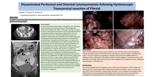
Case report: A 51-year-old perimenopausal lady, on Desogestrel, was referred by the GP for pelvic pain. She had a history of menorrhagia and underwent 2 Hysteroscopic TCRFs 7 years ago. There was no history of open or laparoscopic myomectomy. Ultrasound showed thickened endometrium (10mm) and extrauterine fibroids with uncertain connection to the uterus. She had a palpable pelvic mass and outpatient hysteroscopy which showed a normal endometrial cavity. The diagnostic dilemma prompted an MRI which revealed diffuse leiomyomatosisin the pelvis and left iliac fossa. Her images were discussed in the multidisciplinary team (MDT) meeting and CT was recommended. The CT showed extensive omental nodularity and peritoneal fibroids. This was re-discussed at the MDT and the recommendation was to offer surgical excision and biopsy followed by a hysterectomy. The patient was not keen on surgery and therefore was kept on follow-up. She had a repeat CT 6 months later which showed an increased volume of widespread peritoneal nodularity. Metastatic small bowel gastrointestinal stromal tumour (GIST) was also a differential. The MDT recommended a biopsy as the patient was not keen on major surgery. She was counselled and a mutual agreement to proceed with Laparoscopy and biopsy was made. The laparoscopy showed extensive widespread deposition of fibroid over the entire pelvis. There were adhesions between the uterus, omentumand bowel. A biopsy was taken, and the histology reported Leiomyoma. Further discussion at the MDT recommended complete resection but the patient refused and opted for conservative management.
Discussion: Disseminated peritoneal leiomyomatosis(DPL) is a rare condition that is characterized by nodules of smooth muscle cells located on the peritoneum, omental surface, and abdominal organs. There are mainly two hypotheses: Mullerian theory (De-novo differentiation) vs Iatrogenic theory. Although the distribution of these lesions suggests a metastatic process, DPL generally has a benign clinical course and has been regarded as a metaplastic process. Malignant transformation is extremely rare.
Conclusion: DPL can be misdiagnosed as metastatic cancer, GIST, and mesothelioma. There are few case reports and series, particularly after Laparoscopic fibroid morcellation. This is a unique case where DPL has occurred after a hysteroscopic resection. So it is important to be aware of this differential and to counsel patients thoroughly. At the same time, these cases should be managed by a multidisciplinary team and referred to a tertiary centre if necessary.