1,830 posters, 67 videos, 75 audios, 29 topics, 22 sessions, 4,727 authors, 1,697 institutions
ePostersLive by SciGen Technologies S.A. All rights reserved.
RCOG World Congress 2023
June 12 - 14, 2023 | London, UK

EP.0385
Fibroid migration causing an unusual presentation of postnatal sepsis: A case report.
Poster Presenter
Authors
Affiliations
Part of Topic
Benign Gynaecology
Fibroid migration-an unusual presentation of postnatal sepsis (case report)
Jason Parkash, Yealin Chung, Ida Muslim.
Gloucestershire Hospitals NHS Foundation Trust
Introduction
•Fibroid migration with spontaneous expulsion has been reported post-
partum but is usually a complication of uterine arteryembolisation.
•We present an unusual case of postpartum sepsis, cause by fibroid
migration.
Case Presentation
Background
•28 year old woman, 5 weeks post natal
•emergency caesarean at term for presumed fetal distress at 7cm
dilatation
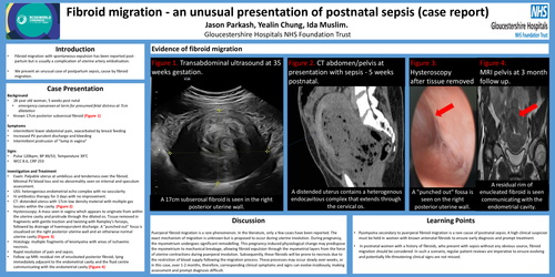
•Known 17cm posteriorsubserosalfibroid(Figure 1)
Symptoms
•intermittent lower abdominal pain, exacerbated by breast feeding
•Increased PV purulent discharge and bleeding
•Intermittent protrusion of ”lump in vagina”
Signs
•Pulse 120bpm, BP 89/53, Temperature 39°C
•WCC 8.6, CRP 253
Investigation and Treatment
•Exam: Palpable uterus at umbilicus and tenderness over the fibroid.
Minimal PV blood loss and no abnormality seen on internal and speculum
assessment.
•USS: heterogenous endometrial echo complex with no vascularity
•IV antibiotics therapy for 3 days with no improvement.
•CT: distended uterus with 17cm low density material with multiple gas
locules within the cavity.(Figure 2)
•Hysteroscopy: A mass seen in vagina which appears to originate from within
the uterine cavity and protrude through the dilatedos. Tissue removed in
fragments with gentle traction and twisting with Rampley’s forceps,
followed by drainage of haemopurulent discharge. A ”punched out” fossa is
visualised on the right posterior uterine wall and an otherwise normal
uterine cavity(Figure 3)
•Histology: multiple fragments of leiomyoma with areas of ischaemic
necrosis.
•Rapid resolution of pain and sepsis.
•Follow up MRI: residual rim of enucleated posterior fibroid, lying
immediately adjacent to the endometrial cavity and the fluid centre
communicating with the endometrial cavity(Figure 4)
Evidence of fibroid migration
Figure1.Transabdominal ultrasound at 35
weeks gestation.
Figure 2.CT abdomen/pelvis at
presentation with sepsis-5 weeks
postnatal.
Figure 3:
Hysteroscopy
aftertissue removed
Discussion
Puerperal fibroid migration is a rare phenomenon. In the literature, only a few cases have been reported. The
exact mechanism of migration is unknown but is proposed to occur during uterine involution. During pregnancy,
the myometrium undergoes significant remodelling. This pregnancy-induced physiological change may predispose
the myometrium to mechanical breakage, allowing fibroid expulsion through the myometrial layers from the force
of uterine contractions during puerperal involution. Subsequently, these fibroids will be prone to necrosis due to
the restriction of blood supply following the migration process. These processes may occur slowly over weeks, or
in this case, over 1-2 months, therefore, corresponding clinical symptoms and signs can evolve insidiously, making
assessment and prompt diagnosis difficult.
Learning Points
•Pyomyomasecondary to puerperal fibroid migration is a rare cause of postnatal sepsis.A high clinical suspicion
must be held in women with known antenatal fibroids to ensure early diagnosis and prompt treatment.
•In postnatal women with a history of fibroids, who present with sepsis without any obvious source, fibroid
migration should be considered. In such a scenario, regular patient reviews are imperative to ensure evolving
and potentially life-threatening clinical signs are not missed.