1,830 posters, 67 videos, 75 audios, 29 topics, 22 sessions, 4,727 authors, 1,697 institutions
ePostersLive by SciGen Technologies S.A. All rights reserved.
RCOG World Congress 2023
June 12 - 14, 2023 | London, UK

EP.0377
A RARE CASE OF MINIMALLY CONJOINED OMPHALOPAGUS TWINS
Poster Presenter
Part of Topic
High Risk Pregnancy
A RARE CASE OF MINIMALLY CONJOINED OMPHALOPAGUS TWINS
Authors –Beena Kumari.B, Mohamed Eraky, Abeer Alzaydai-Obstetrics& Gynecology department, Adan Hospital, Kuwait.
Ahmed E A Hegazi, Fathi A A Yousef, Ahmed A K Hassan, Samer M H Sabbour,Paediatrics department,Adan Hospital ,Kuwait
Introduction
Conjoined twinning is one of the rarest congenital anomalies. According to the site of attachment, every pair of conjoined twins represents a unique event. Minimally conjoined omphalopagus twins (MCOTs) has been recognized as a special subgroup in which omphalopagus twins have union of peritoneal cavities through anterior lower abdominal wall defect with union of distal small intestine and patent urachal structures and associating anorectal malformations. We present this rare case of antenatally undiagnosed minimally conjoined omphalopagus twins to highlight on the challenges faced during delivery & successful resuscitation of twins.
CASE REPORT
A 27 years old Primigravida at 24+6weeks gestation with no relevant medical, surgical or family history and who had conceived spontaneously with Diamniotic Monochorionic twins got admitted on 28/9/2021 with threatened preterm labour. Scan showed both fetuses in breech presentation and adequate liquor in each sac. Patient then received steroids, tractocile and magnesium sulphate for neuroprophylaxis and counseled by neonatologist and obstetrician regarding morbidity, mortality of fetuses and mode of delivery. In 12 days time she progressed to active labour. Emergency Caesarean section was resorted due to fetal distress at 26+5 weeks. During Caesarean section Ɪ twin was extracted as breech, but abdomen was difficult to deliver and stuck near the uterine incision, with gentle traction shoulder and head was delivered. Ɪ Twin still undelivered, ꞮꞮ twin membrane was ruptured and ꞮꞮ twin was extracted as breech and together with Ɪ twin attached ꞮꞮ twin abdomen delivered then shoulder and head. Following delivery both babies were found connected at the umbilicus with thick mass like structure and thick umbilical short cord arising from that mass and running in between the membranes and attached to the centre of placenta .Distance between 2 babies abdomen was around 4 cm. Careful dissection was done identifying 2 umbilical cords ,dissecting in between the 2 cords above 5 cm from the fetal attachment clamped &cut and handed over the babies together attached at the umbilicus to the paediatrician and single placenta with membranes delivered in Toto and caesarean section was completed. This way successfully delivered an undiagnosed omphalopagus conjoined twins.
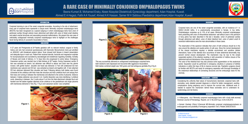
Both newborns were depressed and recovered after aggressive resuscitation. Both babies combined weight was 1600 grams and had ambiguous genitalia.
They underwent emergency separation in 5 hours by the paediatric surgeons and found to have cloacal extrophy and anorectal malformation and common bowel, with both twins having short small bowel and no colon attached at the terminal ileum. Ileostomy and vesicostomy was done. Subsequent follow up showed female karyotype, single introitus with duplicated vagina, one cervix and imperforate anus. Currently both babies are 20 months old, have undergone complete evaluation and multiple follow-up surgeries and being followed up regularly in USA & KUWAIT.
Discussion: Conjoined twins are one of the rarest congenital anomalies, with an incidence of 1 in 50000-1,00,000 births. It is predominantly encountered in female in live births. Omphalopagus comprises up to 10% of all cases. Minimally conjoined omphalopagus twins presenting with union of infraumbilical abdominal wall without union in the perineum or bony pelvis has been described in the last 2 decades. Union of peritoneal cavities through abdominal wall defect, union of distal intestinal tract, union of patent urachi & anorectal malformations were the main abnormalities in these cases. The observation of this spectrum indicates that union of both embryos should be in the area around the allantois and caudal portion of yolk sacs. Since the normal development of the cloacal membrane requires a delicate environment and sensitive cellular interactions, fusion of the allantois &/or occurrence of lower abdominal abnormality may prevent normal cloacal development. The mechanism may be due to the simple suspended position of the allantois resulting in both prevention of midline closure of the abdominal wall and disturbance of the cloacal membrane. The union of the intestinal tract may also produce some augmentation on this anatomical barrier. Separation would be performed in emergency situations in presence of definite indications or within first days of life to improve survival rate.Pediatric surgeons should be familiar with MCOTs and ready for emergency separation with thorough knowledge of the anatomical relationships of connecting structures and the embryologic basis for this anomaly.
Conclusion: Considering the uniformly fatal nature of monochorionic diamniotic conjoined twins with anomalies, early detailed prenatal diagnosis is critical for better management, averting complications during pregnancy and to improve the survival rate. Additional work is needed to explore the mechanism behind these anomalies and to understand its epidemiology and risk factors.