1,830 posters, 67 videos, 75 audios, 29 topics, 22 sessions, 4,727 authors, 1,697 institutions
ePostersLive by SciGen Technologies S.A. All rights reserved.
RCOG World Congress 2023
June 12 - 14, 2023 | London, UK

EP.0349
Acute presentation of unsuspected endometrial stromal sarcoma with uterine rupture and sepsis.
Poster Presenter
Part of Topic
Gynae Oncology
Introduction
·Uterine sarcomas are rare, highly aggressive, gynaecologic malignant tumours.
·Uterine rupture and sepsis are unusual complications.
·Emergency surgery is required on suspicion of acute rupture.
·Only a few reports in the literature describe such an infrequent presentation.
Case Report
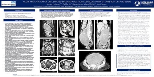
·A 59-year-old woman, P2 (both normal deliveries), postmenopausal for 12 years, with no risk factors had an episode of postmenopausal bleeding. Ultrasound and MRI revealed multiple uterine fibroids with uterus measuring 14.6 x 9.9 x 12 cm and the dominant fundal fibroid 8.2 x 8.2 cm.
·She underwent hysteroscopy which showed atrophic cavity and endometrial biopsy that showed inactive endometrium. She was listed for hysterectomy for multiple fibroids.
·The patient soon noticed that the abdominal mass was rapidly increasing in size, was suffering with severe pain, and losing weight. Multiple attempts were made to access primary care and to expedite surgery.
·Less than 8 weeks later, the patient attended for elective hysterectomy, but was cancelled and patient discharged due to pyrexia.
·The next day she presented with acute abdominal pain, a massively enlarged abdomen sepsis.
·Physical examination revealed pulse 110 bpm, temperature 38°C, distended abdomen with palpable bulky fibroids above the umbilicus with tenderness over the upper abdomen.
·Urgent CT abdomen and pelvis showed a significant increase in the size of the fibroids with overall dimensions of 25 x 24 x 18 cm with a significant intra-abdominal mass effect. The fibroids demonstrated mixed heterogeneous internal densities suggestive of a haemorrhagic component. Small volume and low-density free fluid was also noted within the pelvis.
·Laboratory investigations revealed elevated inflammatory markers (WBC 23, CRP 359, Plt 523), low haemoglobin levels (Hb 101) and coagulation profile abnormalities (INR 2, PT 23).
·Emergency explorative midline laparotomy was undertaken. Intraoperative findings showed a 30cm ruptured multi-fibroid uterus with suspicion of sarcomatous lesions. The tumour was adherent posteriorly to the small bowel and right pelvic side wall. Subtotal hysterectomy, bilateral salpingo-oophorectomy, omental biopsy, and debulking of lesions was carried out. Blood loss was nearly 4L. Five units of RBCs and 2 units of FFP were transfused.
·She was transferred to ICU postoperatively.
·Pathologic analysis demonstrated the uterus to be entirely replaced by high-grade endometrial stromal sarcoma with lymph-vascular space invasion.
·Molecular studies revealed presence of an in-frame fusion transcript involving EPC1 exon 10 and SUZ12 exon 2. A matching fusion transcript of EPC1 and SUZ12 has previously been described as an oncogenic driver in endometrial stromal sarcoma.
·During the weeks post-operatively, the patient continued to deteriorate and again made attempts to access non-emergency care. She was subsequently re-admitted as an emergency.
·CT demonstrated indeterminate lung lesions and pelvic metastatic deposits, causing compression of the right iliac veins and invasion of the right ureter with grade 2 hydronephrosis.
·There was a protracted inpatient stay where the patient developed malignant ileus and required nephrostomy.
·Sadly, her health rapidly deteriorated and her condition was deemed to be palliative. She passed away 2 months after her emergency surgery, and 4 months after initial presentation.
Discussion
·Uterine sarcomas are rare tumours, with incidence 5-7% of all malignant uterine tumours. Endometrial stromal tumours constitute about 0.2% of all uterine malignancies (1,2).
·High-grade endometrial stromal sarcoma became a distinct category in the 2014 World Health Organization (WHO) classification with a mean onset age of 50 years. They are aggressive tumours with quickly spread locally by extension or abdominal implantation. The tumours often present as intrauterine polyp-like bulges or myometrial nodules. The cut section of the tumour shows a fish-flesh appearance, with areas of haemorrhage and necrosis (3).
·The clinical presentation of is similar to that of uterine fibroids, which can present with irregular vaginal bleeding, pelvic masses, and pelvic and abdominal pain.
·Only a few reports in the literature describe cases of haemoperitoneum caused by ruptured uterine sarcomas that initially appeared as benign masses.
·Hysteroscopic-guided biopsy can be useful in diagnosis, particularly where areas of bleeding, necrosis or irregular morphology are noted, but the muscular layer cannot be reached to obtain a biopsy if the mass does not extend into the cavity (5).
·Diagnosis of uterine sarcomas is complex. Clinical features, imaging findings and laboratory investigations can point towards uterine fibroids while overlooking the possibility of uterine sarcomas.
·There are no definitive preoperative diagnostic criteria to distinguish sarcomas from other tumours, and the final diagnosis is usually made after a histopathological examination.
·As an acute presentation of uterine sarcoma and hematoperitoneum is very rare and life-threatening. Emergency surgery to control the bleeding is crucial. While complete resection offers the best chance of long-term survival, following the oncological principles of resection might be challenging as priority goes to achieving haemostasis.
·The mainstay for treatment is surgical. Aggressive tumour debulking is valuable for extrauterine spread. Adjuvant treatment remains a topic of debate. Treatment lines for the advanced disease include systemic chemotherapy, targeted therapy or palliative care (6).
·The rapidly progressive nature of these tumours, as well as the difficulty in diagnosis, and the rarity of these tumours makes it difficult to develop and evaluate new therapies.
Conclusions & Learning Points
·Uterine sarcomas are rare lethal tumours with atypical clinical symptoms and poor prognosis.
·Early identification of uterine sarcomas is critical in improving patient survival and quality of life.
·Acute rupture of uterine sarcoma is a rare complication and risks and benefits of acute surgical management must be carefully considered.
·It is vital that awareness is raised amongst both primary and secondary clinicians so that the possibility of this diagnosis is always at the forefront of the differential diagnosis, particularly in those with rapidly increasing fibroids.
·Any woman with fibroids that appear large or atypical on imaging, should be made aware of the rare possibility of this diagnosis, particularly when deciding upon medical vs surgical management. This will enable the patient themselves to highlight concerns if rapid growth is noted.
·Early referral to regional sarcoma team should be undertaken as soon as diagnosis is suspected so that specialist care can be provided.
·A single point of contact for patients with suspected sarcoma (and their families) is necessary to ensure information is communicated clearly and consistently, and that there are no difficulties in accessing care during what is likely to be an extremely distressing time.
Acknowledgements
We would like to express our heartfelt thanks to the patient and her family, who kindly allowed their personal story to be shared on this platform to raise awareness of this condition.