1,830 posters, 67 videos, 75 audios, 29 topics, 22 sessions, 4,727 authors, 1,697 institutions
ePostersLive by SciGen Technologies S.A. All rights reserved.
RCOG World Congress 2023
June 12 - 14, 2023 | London, UK

EP.0331
Hysteroscopy findings: quadricornuate uterus...?! An unlikely part-congenital, part-iatrogenic uterine anomaly.
Poster Presenter
Authors
Affiliations
Part of Topic
Benign Gynaecology
Background
Congenital uterine anomalies (CUAs) develop when a malfunction occurs during the normal process of Müllerian duct development and fusion. Correctly classifying CUAs is vital to allow accurate counselling regarding reproductive implications and potential management options such as septal resection. The RCOG now recommends 3D transvaginal ultrasound as gold-standard for diagnosis and classification of CUAs, with MRI or laparoscopy and hysteroscopy reserved for complex cases.
We present here a case involving a puzzling uterine anomaly found in a parous woman during diagnostic laparoscopy and hysteroscopy, where the final diagnosis only became apparent following subsequent imaging.
Case Report
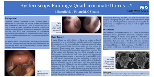
X was a 23-year-old with a history of Caesarean section for breech presentation at term, who had previously been advised of a suspected bicornuate uterus. She presented to the gynaecology clinic with a long history of dysmenorrhoea and menorrhagia, and was booked for diagnostic hysteroscopy and laparoscopy. Examination under anaesthetic demonstrated a longitudinal septum in the proximal third of the vagina, with duplicated cervices. Most unusually, however, hysteroscopic examination via each cervix appeared to demonstrate two distinct cavities separated by a septum (ie. a right and left cavity for each uterus). Laparoscopy was then undertaken as planned: this demonstrated a single uterus which had a slight indent at the fundus but was otherwise normal in appearance with appropriately sited Fallopian tubes. An MRI carried out afterwards provided a definitive diagnosis: X did indeed have a complete septate uterus with duplicated cervices – however her Caesarean section had bisected the distal portion of the intra-uterine septum and healed to leave a defect, which we had been inadvertently traversing during the hysteroscopy, entering the same two cavities via each cervix.
Immediately following her MRI, and just two months after surgery, X fell pregnant again. She underwent cervical length scans in the preterm prevention clinic and is now 32/40; the significance and implications, if any, of such a septal defect remain unclear.
Discussion
This is the first published incidence of a Caesarean section scar healing to leave a permanent defect in a complete uterine septum, although it seems likely given the incidence of Caesarean section that similar cases will exist. It is well documented that even non-obstructive CUAs are associated with menstrual abnormalities: whether X’s septal defect was an incidental finding or whether passage of blood between the two cavities could have worsened her dysmenorrhoea is not known.