1,830 posters, 67 videos, 75 audios, 29 topics, 22 sessions, 4,727 authors, 1,697 institutions
ePostersLive by SciGen Technologies S.A. All rights reserved.
RCOG World Congress 2023
June 12 - 14, 2023 | London, UK

EP.0320
Late miscarriage due to ruptured tubo-ovarian abscess secondary to perforated sigmoid diverticulitis
Poster Presenter
Authors
Affiliations
Part of Topic
High Risk Pregnancy
Audio
Late miscarriage due to ruptured tubo-ovarian abscess secondary to perforated sigmoid diverticulitis
Introduction
Tubo-ovarian abscess (TOA) during pregnancy is a rare occurrence and increases maternal morbidity, mortality and risk of miscarriage. This case follows the rare development of a tubo-ovarian abscess secondary to perforated sigmoid diverticulitis. This led to the outcomes of second trimester miscarriage, maternal sepsis, ICU admission and other life-threatening complications after rupture of the TOA that did not respond to outpatient parenteral antimicrobial therapy (OPAT). The need for multidisciplinary team (MDT) care, early intervention and rigorous management of TOA in pregnancy are highlighted.
Case Presentation
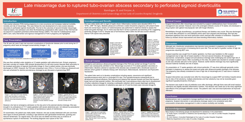
The 30 year old, para 2 lady had recurrent complications of diverticular disease prior to and during her 3rd pregnancy which were all managed conservatively (Images 1 - 4).
She was then admitted under obstetrics at 12 weeks gestation with abdominal pain. Ectopic pregnancy had been outruled at 9 weeks. MRI showed development of a left tubo-ovarian abscess likely secondary to the localised perforation of sigmoid diverticulitis (Image 4). Due to patient wishes, she was eventually discharged on OPAT. However, the TOA was increasing in size (10cm x 11cm) on follow-up ultrasound and she was scheduled for elective percutaneous drainage as per discussion at gynaecology MDT.
However, she had an emergency admission on the day prior to the planned elective drainage. She was brought in by ambulance at 17+1 weeks gestation with severe abdominal pain, not relieved by paracetamol and nitrous oxide gas. There was no history of spontaneous rupture of membranes.
On examination, the patient was vitally stable. However, she was acutely distressed with pain and there were signs of clinical peritonitis, including a rigid abdomen with generalised tenderness, guarding and rebound tenderness. On vaginal exam, the cervix was not dilated and there was no evidence of spontaneous rupture of membranes. The working diagnosis was rupture of the TOA.
Investigations and Results
She was vitally stable with no signs of septic shock. Inflammatory markers were elevated (WCC 17 x 10*9/L, CRP 118 mg/L). Bedside ultrasound confirmed a viable intrauterine pregnancy with present foetal heart and normal liquor volume. CT abdomen/pelvis was performed in maternal interest to assess the bowel and guide drainage. On CT, there was free fluid in all four quadrants and stranding of fat, suggesting peritonitis (Images 5 and 6). Despite lack of full thickness defect within the left tubo-ovarian abscess, interval TOA rupture was presumed.
Clinical Course
Emergency percutaneous ultrasound-guided drainage of the TOA was carried out (Image 7) and the patient was subsequently admitted to ICU. Miscarriage occurred at 17+2 weeks gestation secondary to inflammation. Bacteria cultured on aspirate of the abscess was consistent with a diverticular source of infection which guided IV antibiotic therapy with Vancomycin, Metronidazole, Gentamicin and Piperacillin-Tazobactam.
The patient later went on to develop complications including sepsis, pneumonia and significant hypoalbuminaemia which led to a prolonged ICU stay. The hypoalbuminaemia subsequently led to development of pleural effusions, third-spacing and oedema that required diuresis and ileus that required bowel rest and total parental nutrition. She eventually had 3 percutaneous drains sited due to development of further collections (Image 8), including a subhepatic collection which caused diaphragm splinting. Eventually, interval resolution of collections was seen on CT and the drains were removed (Image 9).
Surgical intervention after miscarriage was excluded by surgical multidisciplinary input. Conservative management was advised. However, due to the prolonged antibiotic course of 10 weeks, she developed a drug eruption rash (global maculopapular rash with fungal infection).
Rehabilitation through physiotherapy, occupational therapy and dietetics was crucial. She was discharged one month after admission on oral antibiotics which were soon stopped post-discharge. She had also developed significant liver and renal dysfunction which resolved after cessation of antibiotics. Outpatient follow-up showed clinical improvement and down-trending inflammatory markers.
Discussion
Although rare, diverticular complications may become more prevalent in pregnancy as incidence of diverticulitis in young women is increasing due to poor diet. This can lead to a greater number of high risk pregnancies requiring MDT care.
Surgical expertise was sought from 9 weeks’ gestation, but there was hesitation to offer surgical management due to high risk of bowel resection in first and second trimesters. All management approaches: conservative, surgical and percutaneous drainage were challenging and carried high risk of miscarriage or preterm labour. After counselling on the risks, the patient was reluctant to consider surgery as she was clinically well prior to the rupture. However, earlier elective drainage may have significantly reduced maternal morbidity and risk of sepsis.
On presentation at 17 weeks gestation with clinical peritonitis, CT was done (although generally contra-indicated in pregnancy) to assess the bowel and guide safe ultrasound-guided drainage of the abscess. The pregnancy was already considered to have a high risk of miscarriage and CT was done in maternal interest.
Surgical intervention was excluded even after the miscarriage by surgical MDT and tertiary hospital advice due to risks of full colostomy, multiple enterotomies and that colostomy may not be reversible given significant hypoalbuminaemia (18-24 g/L).
The decision was made to stop all antibiotics soon after discharge, although there were still small residual collections and slightly elevated inflammatory markers. This decision was made due to the many significant side-effects of the prolonged antibiotic course. The patient’s rash, liver and renal dysfunction all improved afterward.
Conclusion
Multidisciplinary care is essential to manage significant risks and complications posed by TOA in pregnancy. Surgical intervention or percutaneous drainage needs to be considered early after unsuccessful conservative management in order to reduce morbidity and mortality.