1,830 posters, 67 videos, 75 audios, 29 topics, 22 sessions, 4,727 authors, 1,697 institutions
ePostersLive by SciGen Technologies S.A. All rights reserved.
RCOG World Congress 2023
June 12 - 14, 2023 | London, UK

EP.0276
Investigating intrauterine fetal growth after IVF-assisted conception
Poster Presenter
Authors
Charlotte Dunkerley, Lucy Higgins, Reena Perchard, Jack Wilkinson
Affiliations
Part of Topic
Fetal Medicine
Investigating intrauterine fetal growth after IVF-assisted conception
Charlotte Dunkerley1, Reena Perchard2, Jack Wilkinson3, Lucy Higgins4
1Medical Student, University of Manchester; 2Lecturer in Clinical Trial Statistics, University of Manchester;3Academic Clinical Lecturer, University of Manchester; 4Senior Clinical Lecturer in Obstetrics, Manchester University Foundation NHS Trust
Background
Hypothesis and Aims
“In utero growth and birthweight differ by mode of conception”
This study collected pilot data to test feasibility and generate further research hypotheses
Materials and Methods
A prospective IVF-conceiving cohort was recruited to START clinic study (REC: 20/NE/2022). A historic prospectively-studied SC control cohort was used as comparison (NUPS; REC: 10/H1013/9).
Aim 1: BW z-scores were compared between MOC by linear regression
Aim 2: serial ultrasound biometric parameters were measured (BPD, HC, AC, FL, TVol, AVol, 2D/3D EFW).
Ultrasound scan (USS)-determined gestational age was used for all pregnancies.
Serial 2D/3D USS measurements of fetal biometric parameters were plotted to map fetal growth by MOC.
N=131 (SC N=103; fresh ET N= 9; FET N= 20).
All statistical analysis was carried out using Stata/BE (Stata Corp, Texas, USA). P value <0.05 was considered statistically significant and 95% Confidence Intervals (CI) were used.
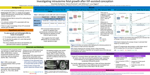
FET-conceived infants were born heavier for gestation than spontaneously conceived infants, even after adjusting for gestation at birth, maternal age and parity. This suggests that birth weight differences are not simply due to longer gestational ages
There was no suggestion of difference in skeletal growth between MOC type (P>0.1; data not shown). Differences in parameters influenced by soft tissue growth between SC and IVF groups were very subtle when graphed. Frozen ET pregnancies tended to lay down soft tissue faster than Fresh ET pregnancies.
Frozen ET pregnancies demonstrate a subtly faster rate of EFW increase than SC and fresh ET pregnancies.
Discussion
Frozen ET conceived infants had higher BW (Table 1) compared to SC peers, even after accounting for gestational age, supporting current literature (3) and suggesting presence of potentially accelerated intrauterine growth.
Our results suggest potential differences in fetal soft tissue deposition may exist. The direction of such differences are inconsistent and limited by a lack of AVol data in the SC cohort.