1,830 posters, 67 videos, 75 audios, 29 topics, 22 sessions, 4,727 authors, 1,697 institutions
ePostersLive by SciGen Technologies S.A. All rights reserved.
RCOG World Congress 2023
June 12 - 14, 2023 | London, UK

PP.0091
LEER in disseminated leiomyomatosis mimicking low-grade sarcoma in a solitary kidney patient
Part of Topic
Gynae Oncology
Objective : Although surgical excision is the treatment of choice for disseminated leiomyomatosis, there has been limited literature addressing the optimum technique and surgical complexity associated with aggressive growth of the tumour, particularly in instances when the diagnosis of benign/malignant disease is unclear.
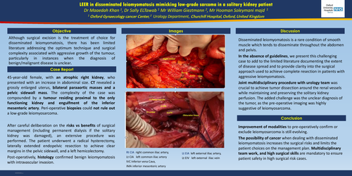
Case report: Our patient is a 45-year-old female, with an atrophic right kidney, who presented with an increase in abdominal size. CT revealed a grossly enlarged uterus, bilateral paraaortic masses and a pelvic sidewall mass. The complexity of the case was compounded by a tumour residing proximal to the only functioning kidney and engulfment of the inferior mesenteric artery. Peri-operative biopsies could not rule out a low-grade leiomyosarcoma. After careful deliberation on the risks vs benefits of surgical management (including permanent dialysis if the solitary kidney was damaged), an extensive procedure was performed. The patient underwent a radical hysterectomy, laterally extended endopelvic resection to achieve clear margins in the pelvic sidewall, and a left hemicolectomy. Post-operatively, histology confirmed benign leiomyomatosis with intravascular invasion.
Discussion: Disseminated leiomyomatosis is a rare condition of smooth muscle which tends to disseminate throughout the abdomen and pelvis. In the absence of guidelines, we present this challenging case to add to the limited literature documenting the extent of disease spread and to provide clarity into the surgical approach used to achieve complete resection in patients with aggressive leiomyomatosis. Joint multidisciplinary procedure with urology team was crucial to achieve tumor dissection around the renal vessels while maintaining and preserving the solitary kidney perfusion. The added challenge was the unclear diagnosis of the tumor, as the pre-operative imaging was highly suggestive of leiomyosarcoma.
Conclusion: Improvement of modalities to pre-operatively confirm or exclude leiomyosarcoma is still evolving. The possibility of cancer when dealing with disseminated leiomyomatosis increases the surgical risks and limits the patient choices on the management plan. Multidisciplinary team work, and high surgical skills are mandatory to ensure patient safety in high surgical risk cases.