1,830 posters, 67 videos, 75 audios, 29 topics, 22 sessions, 4,727 authors, 1,697 institutions
ePostersLive by SciGen Technologies S.A. All rights reserved.
RCOG World Congress 2023
June 12 - 14, 2023 | London, UK

PP.0086
Successful Recovery after Arteriovenous Malformation Excision in Pregnancy Following Occipital Lobe Haemorrhage
Poster Presenter
Authors
Chiran Colombage, Chathura Ratnayake, Sanjaya Athapattu, Sanjeevan Kanagarasa
Affiliations
Part of Topic
High Risk Pregnancy
Successful Recovery after an Arteriovenous Malformation Excision in Pregnancy Following an Occipital Lobe Haemorrhage - A Case Report
Chiran Colombage, Chathura Ratnayake, Sanjaya Athapattu, Sanjeevan Kanagarasa
Teaching Hospital Peradeniya, Sri Lanka
Abstract
Background
Arteriovenous malformation (AVM) is one of the causes of intracranial hemmorrhage, which is a devastating entity in pregnancy. It is rare to have a succesful recovery after an encounter such as this and even rarer to have pregnancy contiued afterwards without a permanat neurological sequale.
Case Description
The case presented here is a 32 year old second para with an uncomplicated and normotensive antenatal period presented with sudden onset bilateral vision loss. CT and MRI brain has confirmed the diagnosis of arteriovenous malformation in the occipital lobe. She underwent a craniotomy and evacuation of haematoma with AVM excision few days after the diagnosis. She made a successful recovery with regard to the vision and other funtionality. Her pregnancy is contuing and foetal condition is satisfactory in terms of foetal growth and well being.
Conclusion
Arteriovenous malformation in pregnancy is a challanging scenario to the patient as well as to the clinicians but can be managed to have a successful neurological and obstetric outcome given the timely and appropriate neurosurgical intervention.
BACKGROUND
Intracranial hemorrhage in pregnancy is a rarely encountered entity which usually has a permanent neurological sequalae as only 20% achieve a meaningful level of functional recovery and 30 day mortality rate accounting for 50%. Underlying cause usually is related to uncontrolled hypertension but other causes such as aneurismal ruptures and arterio-venous malformation are known as well. Timely neurosurgical intervention is necessary but decision to do such is a clinical dilemma since lack of clinical data and proper guidelines related to the scenario.
Here we present a case of a lady who made a successful recovery following a craniotomy and AVM excision in early second trimester following an intracranial hemorrhage in occipital lobe.
CASE DESCRIPTION
A 32 year old in her second pregnancy, 13 weeks into her pregnancy admitted to the preliminary care unit with sudden onset of bilateral vision loss and confusion which was superseded by two days of headache. She has been apparently well in her pre-pregnancy years and had an uncomplicated and normotensive antenatal period apart from one admission of excessive vomiting at 12 weeks of gestation, which she has recovered with oral antiemetics.
At her presentation to the preliminary care unit, her Glasgow Coma Scale was 14/15 and the neurological examination showed normal upper, lower limb examination and intact cranial nerves except the optic nerve. Papilledema was not noted in fundoscopy. Blood pressure was 130/70, pulse rate 68 bpm, reflexes were normal with no clonus noted. Bedside ultrasound scan showed a viable foetus.
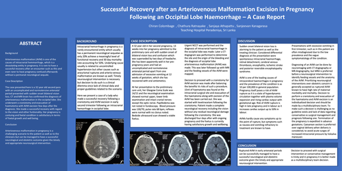
Urgent NCCT was performed and the diagnosis of intracranial haemorrhage in the occipital lobe was made. Later a CT- Angiogram was performed to determine the site and the origin of the bleeding and the diagnosis of occipital lobe arteriovenous malformation (AVM) was made. This was later followed up with MRI and the feeding vessels of the AVM were mapped.
Decision to proceed with a craniotomy for AVM excision was made after balancing the benefits Vs risks of such a procedure. 15ml of haematoma was found at the intracranial surgical site and evacuation of the haematoma along with excision of the AVM has been carried out. She was started with levetiracetam following the craniotomy. Patient made a complete neurological recovery including the vision without any residual neurological damage following the craniotomy. She was discharged four days after with ongoing pregnancy and the foetus is currently having satisfactory growth and wellbeing.
DISCUSSION
Sudden onset bilateral vision loss is alarming to the patient as well as the treating clinician. Considered differential diagnosis at the presentation were spontaneous intracranial haemorrhage, retinal detachment, cerebral venous thrombosis, encephalitis, ischemic stroke and posterior reversible encephalopathy syndrome.
AVM is one of the leading causes of intracerebral haemorrhages in pregnancy and the prevalence of the condition is as 10 per 100,000 in general population. Pregnancy itself poses a risk of AVM rupture as it is a state of hyperdynamic circulation together with plasma volume expansion and rising cardiac output with gestational age. Risk of AVM rupture is high in late pregnancy and in labour as it increases cardiac output up to 50% in labour.
AVMs hardly cause any symptoms up to the point of rupture, but symptoms such as nausea and vomiting refractory to treatment are known to have.
Presentations with excessive vomiting in first trimester, such as in this patient are often misdiagnosed due to the low prevalence and the vague symptomatology of the condition.
Diagnosing of an AVM can be done by neuroimaging with CT-Angiography or MR-Angiography, but MRA is preferred before a neurosurgical intervention to identify feeding vessels and the anatomy of the AVM. Prioritizing neurosurgical interventions over obstetric concern is generally accepted as ruptured AVM known to have high rate of maternal morbidity and mortality. Decision to perform a craniotomy and evacuation of the haematoma and AVM excision is an individualized decision and should be made by a multidisciplinary team. To make such a decision is challenging as no guideline exists and lack of data regarding conservative vs surgical management and prognosis following one. Termination of the pregnancy is expedited in advance gestations. Caesarean section is preferred over vaginal delivery when delivery is considered, to avoid acute surges of increased intracranial pressure by Valsalva maneuver.
CONCLUSION
Ruptured AVM in early antenatal periods can be successfully managed to have a successful neurological and obstetric outcome given the timely and appropriate neurosurgical intervention. Decision to proceed with surgical intervention or conservative management is tricky and in pregnancy it is better made as a multidisciplinary team decision.