1,830 posters, 67 videos, 75 audios, 29 topics, 22 sessions, 4,727 authors, 1,697 institutions
ePostersLive by SciGen Technologies S.A. All rights reserved.
RCOG World Congress 2023
June 12 - 14, 2023 | London, UK

EP.1646
Uterine carcinosarcoma with vaginal skip lesion;A case report
Poster Presenter
Part of Topic
Gynae Oncology
Introduction
Uterine carcinosarcoma (UCS) is a rare extremely aggressive undifferentiated metaplastic carcinoma that accounts for less than 5% of all uterine malignancies. UCS is previously known as malignant mixed Müllerian tumors because they comprise both carcinomatous and sarcomatous elements. UCS origin from a single malignant epithelial clone and is more comparable to endometrial adenocarcinoma than uterine sarcoma. International Federation of Gynecology and Obstetrics (FIGO), classified UCS as a non-endometrioid tumor with high-grade behavior. UCS is staged using the FIGO staging classification for cancer of the corpus uteri which is the best independent predictor of the outcome. Unfortunately at presentation, 10% of patient have distant metastasis and 45 % has extrauterine spread. Thus prognosis is often poor and the five-year survival rate is approximately 30%. In this study, we present a rare case of uterine UCS with a vaginal skip lesion.
Case report
A 64‑year‑old post-menopausal lady presented to a tertiary care hospital in November 2021 with post-menopausal bleeding. She reached menopause at the age of 48 years and was not on hormone replacement therapy. She is a mother of three, all delivered vaginally, and was treated for hypertension and diabetes mellitus. She doesn’t have a significant past gynecological history or family history related to malignancies. She had four episodes of mild vaginal bleeding lasting for a one-day duration with spontaneous settlement over the past year. Her blood pressure was 130/90mmHg and general, cardiovascular, and respiratory examination was normal. No vaginal or cervical pathology was identified in the speculum examination but active bleeding was noted. Bimanual vaginal examination revealed an anteverted mobile non-tender uterus size comparable to a 12-week gravid uterus. Basic investigations were normal. The initial ultrasound scan (USS) of the abdomen and pelvis showed an enlarged uterus with an endometrial thickness of 6.3mm. Bilateral ovaries appeared atrophic and no evidence of significant peritoneal fluid was noted. Histology of the endometrial sample revealed endometrial carcinoma with an associated squamous component. Unfortunately the patient was lost to follow-up due to the COVID-19 pandemic.
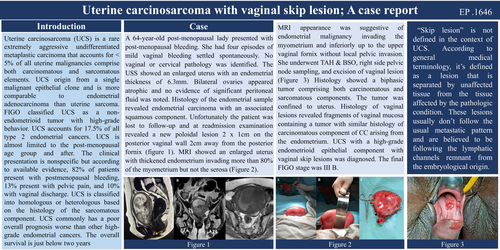
The patient was readmitted to the same unit in May 2022 when she was found to have a new poloidal lesion 2 x 1cm on the posterior vaginal wall 2cm away from the posterior fornix (figure 1). This lesion was suspected to be a vaginal skip lesion. Cervix appeared healthy and the uterus has enlarged compatible with a 16-week gravid uterus. Otherwise, general examination and basic investigations were normal. She underwent an MRI abdomen and pelvis. MRI showed an enlarged uterus measuring 8cm x 11.2cm x 10cm (Figure 2) with thickened endometrium invading more than 80% of the myometrium. No invasion was seen beyond the uterine serosa. Inferiorly the lesion extended involving the cervix and the right lateral and posterior fornices of the upper vagina. Both ovaries were adhered to the anterior uterine wall and the left external iliac lymph node was enlarged. The bladder and rectum appeared normal and no free fluid was noted. The bones of the imaged region appeared unremarkable. MRI appearance was suggestive of endometrial malignancy invading the myometrium and inferiorly up to the upper vaginal fornix without local pelvic invasion beyond the uterine serosa.
She was transferred to the National Cancer Institute for further management. Examination under anesthesia revealed 3 vaginal lesions 3, 6, and 9o clock positions measuring 2 x 2 cm, and uterine growth protruding through the external cervical OS. A Midline laparotomy was performed under general anesthesia. The uterus was bulky and compatible with a 16-week gravid uterus without features of serosal invasion (Figure 3). Bilateral fallopian tubes and ovaries appeared normal. An enlarged right side pelvic lymph node was noted and the omentum appeared normal. She underwent total abdominal hysterectomy with bilateral salpingo-oophorectomy, right side pelvic node sampling, and excision of vaginal lesions.
Macroscopically, the cut surface of the uterus showed a white color lesion occupying the entire endometrium involving the outer half of the myometrium. Microscopically, sections taken from the uterus showed a biphasic tumor comprising both carcinomatous and sarcomatous components. The tumor involved 80% myometrium and both ectocervix and endocervix were involved with 20% endocervical stromal involvement. The tumor is 1 mm away from the ectocervical resection margin. Bilateral parametria were free of the tumor. Bilateral fallopian tubes and ovaries were unremarkable. Omental fatty tissue and Right pelvic lymph nodes were devoid of tumor deposits.
Histology of vaginal lesions revealed fragments of vaginal mucosa containing a tumor with similar histology of carcinomatous component of CC arising from the endometrium. Considering the immune profile, estrogen receptors (ER) were positive in glandular areas but negative in sarcomatous areas. Both glandular and sarcomatous components were negative for carcinoembryonic antigen (CEA) but strongly positive for vimentin staining. UCS with a high-grade endometrioid epithelial component with vaginal skip lesions was diagnosed. The final FIGO stage was III B.
Discussion
During the last several decades the population incidence of UCS has increased. In 1980 the incidence was 1.7% and in 2020 it has increased to 5.6%. CS accounts for 17.5% of all type 2 endometrial cancers. UCS is almost limited to the post-menopausal age group and after 70 years the incidence begins to increase further. Recently there has been an increasing incidence among women aged 60 to 69 years. The clinical presentation is nonspecific but according to available evidence, 82% of patients present with postmenopausal bleeding, 13% present with pelvic pain, and 10% with vaginal discharge.
Traditionally endometrial cancer is classified into estrogen-dependent Type I cancer and relatively estrogen-independent type II cancer. UCS is considered a type II cancer. Recently it has been proposed to classify UCS according to genetic profile following the findings of the Cancer Genome Atlas (TCGA) database. Thus endometrial cancers are classified into four subtypes which are POLE-ultramutated, MSI hypermutated (MSI-H), copy number (CN)-low, and copy number (CN)-high. The frequency of genetic subtypes differs among studies. According to a recent meta-analysis, this classification has shown promise in predicting the survival outcome.
UCS is classified into homologous or heterologous based on the histology of the sarcomatous component. There is no common form, both types are seen in equal frequency. In both types, the carcinomatous component is an adenocarcinoma. It can be either endometrioid, serous, or clear‑cell type. In the homologous type, the sarcomatous component can be a fibrosarcoma, leiomyosarcoma, or an endometrial stromal tumor. The sarcomatous component is made up of tissues that are non‑native to the uterus in heterologous tumors. These include osteosarcoma, rhabdomyosarcoma, liposarcoma, and chondrosarcoma. Both carcinomatous and sarcomatous elements of UCS appear to have a similar mutational profile with varying frequencies. According to recent molecular studies, both elements were found to be originating from carcinoma lineage. The sarcomatous elements dedifferentiate subsequently.
When considering the staging almost half of the cases are stage I and the other half are advanced-stage cancer. According to available statistics 43.9% of cases are diagnosed at stage I, 8.7% at stage II, 22.9% at stage III, and 24.4% at stage IV. There has been a stage shift identified during the past several decades from advanced to early stage and an increase in identifying lymph node metastasis. This is believed to be due to more efficient lymphadenectomy and early detection of cancer due to endometrial sampling for postmenopausal bleeding.
UCS commonly has a poor overall prognosis worse than other high-grade endometrial cancers. The overall survival is just below two years and stage-specific 5-year survival rates are 54.8%, 36.9%, 24.9%, and 9.2% from stage I to stage IV disease respectively. Survival of UCS depends on several factors. Similar to other endometrial cancers large tumor size (≥5 cm), deep myometrial invasion, lymphovascular space invasion, malignant cells in peritoneal fluid, nodal involvement, and distant metastases are poor prognostic factors. Apart from the above sarcoma dominance is an independent poor prognostic factor. Poorest survival is seen in tumors with a combination of high-grade carcinoma and heterologous sarcoma. The carcinoma element tends to metastasize to distant sites and the sarcoma elements show locoregional spread. The carcinoma component tends to metastasize to pelvic and para-aortic nodes while sarcoma tends to spread to the vagina, cervical trauma, and cervical stroma. Commonly both types’ metastases to the omentum
Although “skip lesion” is not defined in the context of uterine carcinoma, according to general medical terminology, it’s defined as a lesion that is separated by unaffected tissue from the tissue affected by the pathologic condition. These lesions are described in relation to metastatic carcinoma, polyarteritis nodosa, and Crohn’s disease. These lesions usually don’t follow the usual metastatic pattern and are believed to be following the lymphatic channels remnant from the embryological origin. Skip lesions are not defined as related to UCS and no previous studies were found.
UCS is treated as a high-grade endometrial cancer. According to recent evidence, combined multimodality treatment of surgery, chemotherapy, and radiotherapy, improves survival outcomes. The effect is more evident in stage I disease. Surgery has been the mainstay treatment. As for all gynecological malignancies, the aim is complete cytoreduction. But in advanced UCS where this is difficult even the optimal debulking with a residual tumor of ≤1 cm is associated with improved survival. Because of the high chance of microscopic lymph node metastasis, hysterectomy-based surgery with lymphadenectomy is recommended even for early-stage disease. During the last, three decades, the utilization and the quality of lymphadenectomy have also improved
For advanced-stage disease use of chemotherapy and vaginal brachytherapy has increased during the last 30 years. In 2016 almost two-thirds of women with UCS received chemotherapy. Platinum-based agents (carboplatin and cisplatin), alkylating agents (ifosfamide), and paclitaxel have been shown to be useful for UCS. Few recent studies have evaluated the possibility of histologic pattern-specific chemotherapy which is under evaluation. Because the sarcomatous element is the most important factor that determines loco-regional tumor spread, for tumors with sarcoma dominance adding radiotherapy to chemotherapy improves the survival rates. With the increased use of brachytherapy, the use of external beam radiotherapy has been reduced. An increase in the utilization of lymphadenectomy may be the reason. Vaginal brachytherapy is found to be more effective compared to external beam radiotherapy.