1,830 posters, 67 videos, 75 audios, 29 topics, 22 sessions, 4,727 authors, 1,697 institutions
ePostersLive by SciGen Technologies S.A. All rights reserved.
RCOG World Congress 2023
June 12 - 14, 2023 | London, UK

EP.1639
The most unusual presentation of Ovarian Dermoid Cyst so far
Poster Presenter
Authors
Affiliations
Part of Topic
Benign Gynaecology
EP.1639
The most unusual presentation of Ovarian Dermoid Cyst so far
Das,R;Naik,S; Heera P
Max Super Speciality Hospital, Saket, Delhi, India
Introduction The most common type of benign germ cell tumor is Dermoid cyst (Mature cystic teratoma), mostly diagnosed with Ultrasonography or MRI. We report a case of large left ovarian dermoid cyst which on course of laparoscopic removal, was found to be not attached with the ovary but lying separately as a cystic mass thus raising the question of its origin. This has been the most unusual presentation we have learned so far that prompted us to report this case.
Objective: To identify the unusual and rare presentations of Dermoid cyst thus alleviate in accurate diagnosis
Case report: A 41 year old P2L2 came with c/o pain in abdomen on & off for 1 month with her LMP on 24/8/2022 with regular cycle. On PV examination a boggy globular mass palpable of 4*3cm, mobile and tender at left fornix. Her USG reported a solid cystic lesion, 50*42*24mm with echogenic area & multiple air fluid level closely abutting the left ovary. MRI reported a left paraovarian, multiseptate cystic lesion measuring 49*25*41mm, S/O left adnexal dermoid. The tumor markers were normal.
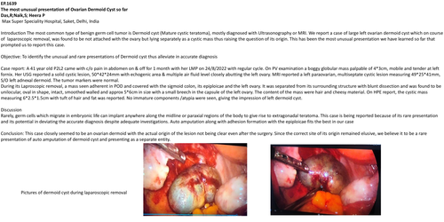
During its Laproscopic removal, a mass seen adherent in POD and covered with the sigmoid colon, its epiploicae and the left ovary. It was separated from its surrounding structure with blunt dissection and was found to be unilocular, oval in shape, intact, smoothed walled and approx 5*6cm in size with a small breech in the capsule of the left ovary. The content of the mass were hair and cheesy material. On HPE report, the cystic mass measuring 6*2.5*1.5cm with tuft of hair and fat was reported. No immature components /atypia were seen, giving the impression of left dermoid cyst.
Discussion
Rarely, germ cells which migrate in embryonic life can implant anywhere along the midline or paraxial regions of the body to give rise to extragonadal teratoma. This case is being reported because of its rare presentation and its potential in deviating the accurate diagnosis despite adequate investigations. Auto amputation along with adhesion formation with the epiploicae fits the best in our case
Conclusion: This case closely seemed to be an ovarian dermoid with the actual origin of the lesion not being clear even after the surgery. Since the correct site of its origin remained elusive, we believe it to be a rare presentation of auto amputation of dermoid cyst and presenting as a separate entity.