1,830 posters, 67 videos, 75 audios, 29 topics, 22 sessions, 4,727 authors, 1,697 institutions
ePostersLive by SciGen Technologies S.A. All rights reserved.
RCOG World Congress 2023
June 12 - 14, 2023 | London, UK

EP.1619
Delayed presentation of large bowel injury following laparoscopic hysterectomy and bilateral salpingectomy
Poster Presenter
Part of Topic
Benign Gynaecology
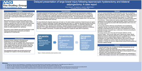
Delayed presentation of large bowel injury following laparoscopic hysterectomy and bilateral salpingectomy: A case report
Introduction
Bowel injury during laparoscopic gynaecological surgery is a rare and potentially life-threatening complication. The incidence of bowel injury in laparoscopic surgery is 0.13% with the most common site of injury being the small bowel followed by the large bowel.
Most bowel injuries occur during Veress needle and trocar insertion. The incidence of injuries caused by Veress needle insertion is reported to be 0.23% out of which 2.8% were bowel injuries. Electrosurgical injuries are the next most common cause with a significant proportion of up to 40% of thermal injuries not being recognised intra- operatively.
We present a case of delayed recto-sigmoid perforation a week following total laparoscopic hysterectomy and bilateral salpingectomy for endometriosis.
Case Presentation
39-year-old woman underwent a total laparoscopic hysterectomy (TLH) and bilateral salpingectomy. Veress needle was used for insufflation with a closed technique used for primary access. All ports were inserted under direct vision. Surgical instruments used were Harmonic scalpel and bipolar diathermy. The operation was uneventful and bowel injury was not identified intra-operatively. Post-operatively the patient was recovering well and was discharged from hospital on the first post-operative day.
The patient presented the following day to the emergency department with peritonitis and haemodynamic instability. A computer tomography (CT) scan has shown a small 3.2x2.6x3.2cm collection in the right adnexa and a 3.4x 4.4x2.4cm collection at the vaginal vault. A diagnostic laparoscopy was done showing a fibrinous tissue on the rectum and sigmoid colon and purulent pelvic fluid which was washed out with insertion of a drain . No bowel perforation was identified. The patient was subsequently transferred to the intensive care unit (ITU) and was subsequently stepped down to the ward when her inflammatory markers were improving.
Five days following the diagnostic laparoscopy and wash out, the patient started complaining of abdominal pain and vaginal bleeding. On speculum examination, feculent vaginal discharge was noted. A CT abdomen was performed which has shown a large pelvic faecally contaminated extra-luminal collection implying bowel perforation. An exploratory laparotomy was done identifying a recto- sigmoid junction perforation. Hartman’s procedure was performed and a washout for faecal peritonitis. A 1cm vaginal vault dehiscence was identified. Post-operatively the patient developed dehiscence of colostomy which was managed conservatively. The patient was discharged from hospital two weeks after the laparotomy.
Discussion
This is an unusual presentation of bowel perforation which was not identified in the diagnostic laparoscopy following the primary operation. Approximately 75% of thermal injures to the bowel are unrecognised at the time of surgery. Symptoms of bowel perforation following electrothermal injury present 4 to 10 days post-operatively whereas in cases of traumatic perforation the symptoms present within 12 to 36 hours. 1,2 Direct contact burn injuries can generally be identified intra-operatively and can therefore be treated appropriately. However conductive burns are usually not recognised and present after bowel perforation whichoccurs one to two weeks later.3,4
The time delay between initial injury and perforation can be explained by coagulation necrosis. Intra-operative findings for delayed bowel perforation secondary to electrothermal injury are the same as the appearances for bowel perforation becauseof traumatic injury: a white area of necrosis surrounding the perforation is usually seen.3
The energy devices used in this case are the bipolar diathermy and the harmonic scalpel . They are both widely used in laparoscopic surgery. Bipolar diathermy functions by delivering electrical current to coagulate and cut tissues, whereas the harmonic scalpel employs ultrasonic vibrations for tissue disintegration and vessel sealing. Bipolar diathermy offers precise cutting and coagulation capabilities but carries a higher risk of thermal injury due to heat generation. In contrast, the harmonic scalpel exhibits reduced thermal spread and minimized collateral damage.
Conclusion