1,830 posters, 67 videos, 75 audios, 29 topics, 22 sessions, 4,727 authors, 1,697 institutions
ePostersLive by SciGen Technologies S.A. All rights reserved.
RCOG World Congress 2023
June 12 - 14, 2023 | London, UK

EP.1606
Ovarian Vein Thrombosis Post-Hysterectomy: a literature review of incidence, implications, and management.
Poster Presenter
Authors
Affiliations
Part of Topic
Benign Gynaecology
Ovarian Vein Thrombosis Post-Hysterectomy
A literature review of risk factors, incidence, implications, and management
Hallikeri. A, Vaselli. N, M, Walker. G
NTRODUCTION
Ovarian vein thrombosis (OVT) is rare type of venous thromboembolism, that occurs in 0.05-0.18% of vaginal births and in 2% of caesarean deliveries(1), however its prevalence post hysterectomy is still widely debated. This literature review aims to discuss risk factors, implications and management of this venous thromboembolic event.
METHOD
A literature review was conducted on PubMed and Google Scholar. The search terms ‘ovarian vein thrombosis’, ‘hysterectomy complications’ and ‘venous thrombosis’ were used to identify peer-reviewed and original articles published in English between 1990 – 2022. Articles were analysed and literature was categorised according to clinical presentation, investigation, prevalence, complications and management.
PATHOPHYSIOLOGY
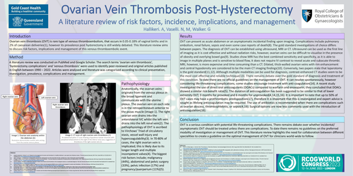
Anatomically, the ovarian veins originate from the venous plexus in the broad ligament and communicate with the uterine plexus. The ovarian vein on each side is in the retroperitoneum anterior to the psoas muscle (image 1). The right ovarian vein drains into the anterolateral IVC whilst the left vein drains into the left renal vein(2). The pathophysiology of OVT is ascribed to Virchows’ Triad of circulatory stasis, vessel wall injury and hypercoagulability(3). In 70-80% of cases, the right ovarian vein is implicated; this is likely due to its longer length and multiple incompetent valves(4). The biggest risk factors include; malignancy (44%), abdominal and pelvic surgery (33%), pelvic infection (17%) and pregnancy/puerperium (11%)(5).
RESULTS
OVT can present as acute abdomen or an asymptomatic incidental finding upon imaging. Complications include pulmonary embolism, renal failure, sepsis and even some case reports of death(8). The gold standard investigations of choice differs between papers. The diagnosis of OVT can be established using ultrasound, MRI or CT. Ultrasound can be used as the first line of imaging as it is cost effective and without radiation risks, however, ovarian veins can be difficult to visualise in the presence of obesity and overlying bowel gas(9). Studies show MRI has the best combined sensitivity and specificity, as it is able to image in multiple planes and is sensitive to blood flow, it does not require IV contrast to reveal acute and subacute thrombi. MRI, however, is more expensive and time consuming than a CT. Dilated, thick-walled ovarian veins with rim enhancement and central hypodensity are considered to be the main CT imaging findings(10). Conversely, two papers state that laparotomy is the gold standard(11,12). Although multiple modalities can be used for diagnosis, contrast-enhanced CT studies seem to be the most cost effective and reliable technique(13). There remains debate over the gold standard of diagnosis and treatment of this condition. To date there are no official guidelines on the management of OVT. It can resolve spontaneously, however considering life-threatening complications, some studies encourage treatment with anti-coagulation(14). A recent study investigated the use of direct oral anticoagulants (DOACs) compared to warfarin and enoxaparin; they concluded that DOACs showed a similar risk-benefit ratio(5). The duration of anticoagulation has been suggested to be similar to that of lower extremity DVT; 3 months for provoked and 6 months for unprovoked(4,14,15,16). It is important to note that up to 50% of OVT cases may have a prothrombotic predisposition(17), therefore it is important that this is investigated and expert advice is sought as lifelong anticoagulation may be required. The use of antibiotics is recommended when there are complications such as ovarian abscess, thrombophlebitis, or sepsis(4,16). Surgical options are now less commonly used with the introduction of anticoagulation(16).
CONCLUSION
OVT is a serious condition with potential life-threatening complications. There remains debate over whether incidental/ asymptomatic OVT should be treated unless there are complications. To date there remains no guidelines on the preferred modality of investigation or management of OVT. This literature review highlights the need for collaboration between different specialities to create a guideline on the optimal management of OVT for clinicians world-wide to follow.