1,830 posters, 67 videos, 75 audios, 29 topics, 22 sessions, 4,727 authors, 1,697 institutions
ePostersLive by SciGen Technologies S.A. All rights reserved.
RCOG World Congress 2023
June 12 - 14, 2023 | London, UK

EP.1574
PRIMITIVE FOREGUT MALFORMATION- BRONCHOGENIC CYST ANTENATAL AND POSTNATAL MANAGEMENT-A CASE REPORT
Poster Presenter
Authors
Affiliations
Part of Topic
Fetal Medicine
INTRODUCTION
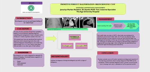
Bronchogenic cysts are rare and benign budding of the ventral foregut
CASE REPORT
30-years-oldprimi-gravida,referredfrom a periphery hospital for second opinion regarding diagnosis, counselling and management of mass in thoracic cavity of fetus at 21 weeks of gestation around 2.6x2.1cm.
We repeated scan from our specialized fetal medicine department which showed single alive fetus with suspected diagnosis of bronchogenic cyst in right lung approximately 32.6 x 23.7 mm, with no other gross fetal anomalies or any other signs of fetal hydrops
DIAGNOSIS
Bronchogenic cyst is easily diagnosed on antenatal 2D ultrasound imaging. Ultrasound features suggestive of bronchogenic cyst are
Post-natallybaby was taken to NICU, where baby was maintained his saturations and remained vitally stable. So CT scan was planned, which showed well defin ed cystic structure within the right lung field.Baby was discharged and was kept on follow up with the plan of surgery at six months of life. At the age of 5 months came in emergency with presenting complaint of progressively worsening cough. After workup baby was admitted and was planned for right bronchoscopy and thoracotomy.
HISTOPATHOLOGY:
confirms the diagnosis of benign bronchogenic cyst with no signs of malignancy
CONCLUSION
Routine anomaly scan at 20 weeks’ gestation can diagnosed majority of the fetal lung anomalies, but for proper diagnosis Fetal MRI can be considered. Timely antenatal diagnosis helps in proper counseling and how to manage such case in antenatal as well as postnatal period