1,830 posters, 67 videos, 75 audios, 29 topics, 22 sessions, 4,727 authors, 1,697 institutions
ePostersLive by SciGen Technologies S.A. All rights reserved.
RCOG World Congress 2023
June 12 - 14, 2023 | London, UK

EP.1572
Atypical radiological appearance of prolapsing degenerated fibroid with acute haemorrhagic presentation
Poster Presenter
Authors
AISHA RAZZAQUE, OMAR HEGAZY, DINA AL DEWAGI, MANON VAN SETERS
Affiliations
Part of Topic
Benign Gynaecology
Title:
Atypical radiological appearance of prolapsing degenerated fibroid with acute hemorrhagic presentation.
Objective:
This case report highlights the unusual presentation with diagnostic difficulties of degenerated fibroid.
Case Report:
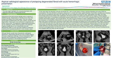
In this case report we describe a 46-year-old female P2+1 with a 2-year history of heavy menstrual bleeding and using COCP for contraception. She was admitted to emergency department in a shock with heavy vaginal bleeding. Examination revealed active bleeding with clots and a prolapsing mass within the vagina with the cervix dilated encircling the mass. Haemoglobin was 48g/L and major hemorrhage protocol was initiated. After stabilization with blood products, the patient was transferred for imaging. Ultrasonography showed a cystic lesion in the posterior endometrial mucosa measuring 8.4 x 3.6 x 3.67 cm with a possible 3mm tract on its inferior part possibly hemorrhagic polypoid / sub mucosal fibroid. The cystic structure showed no classic radiological findings of fibroid, which gave the impression of a possibility of bleed within the cystic structure. MRI was carried out and reported 8 cm pear shaped polypoid bloodfilled cystic lesion extending from myometrial fundal region down protruding and dilating cervix and prolapsing within vagina. Examination under anesthesia was done with opening the cystic structure to reveal 50mls of blood followed by hysteroscopy, which demonstrated a small cavity, and attempts to pass the scope around the mass within the cervix were unsuccessful. Partial resection of the mass with needle diathermy took place and the mass had firm texture and showed classic shape of fibroid in layers. Histopathology reported leiomyoma with degeneration and fibrinoid necrosis. The patient received six units of blood. GnRHa injection was given postoperatively, and outpatient MRI and follow up was arranged.
Case Discussion:
Leiomyoma is the most common benign tumor in women of childbearing age. Degeneration of fibroids occurs when they enlarge and outgrow their blood supply. They can undergo several types of degeneration like cystic, hyaline, myxoid and red degeneration. This is seen more commonly in pregnancy and women using COCP. The diagnosis of degenerating leiomyoma can be challenging. Whilst ultrasonography is the preferred imaging modality due to non-invasiveness and cost effectiveness, MRI is a more accurate modality for detecting, localizing and characterizing fibroid in more complex cases.
Conclusion:
Although fibroids usually have characteristic radiological appearance, degenerating fibroids can have variable patterns and pose a diagnostic challenge. Clinical interpretation, MDT approach and consideration of atypical presentation is of paramount importance in life threatening events.
Categories:
Benign Gynecology.
Authors and Affiliations:
Aisha Razzaque (Presenting) Clinical Fellow Obstetrics & Gynecology Worcestershire Acute Hospitals NHS Trust
Omar Hegazy Clinical Fellow Obstetrics & Gynecology Worcestershire Acute Hospitals NHS Trust
Dina Al Dewagi Consultant Radiologist Worcestershire Acute Hospitals NHS Trust
Manon Van Seters Consultant Obstetrics & Gynaecology Worcestershire Acute Hospitals NHS Trust