1,830 posters, 67 videos, 75 audios, 29 topics, 22 sessions, 4,727 authors, 1,697 institutions
ePostersLive by SciGen Technologies S.A. All rights reserved.
RCOG World Congress 2023
June 12 - 14, 2023 | London, UK

EP.1558
An unusual membranous lesion, which mimics fibroid or polyp found at Hysteroscopy.
Poster Presenter
Authors
Tharaka Nanayakkara, Kumara Dissanayake, Sanjaya Walawenayaka, Chanaka Palansooriya
Affiliations
Part of Topic
Benign Gynaecology
An unusual membranous lesion, which mimics fibroid or polyp found at Hysteroscopy.
Sandaruwan NKTS1, Dissanayaka MAK1, Walawe Nayaka S1, Palansooriya PMCK1
1 Teaching Hospital, Anuradhapura, Srl Lanka
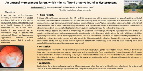
It was not easy to find literature discussing a lesion which is a whitish membrane bulging in to the uterine cavity which mimics intra uterine polyp or a fibroid. However, it is only a membrane but not a solid lesion. Initially it was assumed as an endometrial polyp or pedunculated submucosal fibroid by hysteroscopic appearance, however further evaluation shown that it is only a membrane.
A 65-year-old multiparous woman with DM, HTN and BA was presented with a postmenopausal per vaginal spotting and initial ultrasound revealed thickened endometrium. Further assessment by pelvic ultrasound suggested it as a pedunculated fibroid or as an endometrial polyp. Speculum examination revealed a normal looking cervix. Vaginal examination was unremarkable with normal sized anteverted uterus. Pelvic ultrasonography confirmed a 7cm by 4cm Uterus with an endometrial thickness of 25mm without adnexal masses. Small echogenic fluid collection of 2mm by 3mm was found inside the endometrial thickening.
With the findings decided to perform a Hysteroscopy +/- Dilatation and Curettage. During the Hysteroscopy it was difficult to visualize the bilateral osteas and the upper part of the endometrial cavity. There was a bulging in to the cavity which was mimicking a polyp or pedunculated fibroid. its lining epithelium was similar to a membrane. Therefor it has been decided to proceed for D & C carefully to evaluate the cavity contour and take sample for histopathological evaluation. Repeated hysteroscopy visualized the entire uterine cavity with B/L osteas without significant abnormality. Interestingly there were no such curetting other than blood/fluid/mucus like tissues. Histo-pathology was normal.
The endometrium consists of a simple columnar epithelium, numerous tubular glands, supported by vascular stroma. It divided in to three layers: stratum compactum, stratum spongiosum and stratum basalis. Other than Fibroids, Polyps description of soft tissue mass covered with a membrane mimicking to a polyp or pedunculated fibroid was not found in the literature. Common causes for the thickening of the endometrium or bulging to the cavity are endometrial polyps, endometrial hyperplasia, adhesions or pedunculated fibroids.
Bulging in to the endometrial cavity may be a different pathology other than polyp or fibroids. So, evaluation of the endometrial cavity in AUB with hysteroscopy is highly informative comparison with pelvic ultrasound alone in certain cases.