1,830 posters, 67 videos, 75 audios, 29 topics, 22 sessions, 4,727 authors, 1,697 institutions
ePostersLive by SciGen Technologies S.A. All rights reserved.
RCOG World Congress 2023
June 12 - 14, 2023 | London, UK

EP.1544
A RARE CASE OF COLLISION TUMOR OF THE OVARY
Poster Presenter
Authors
Affiliations
Part of Topic
Benign Gynaecology
Audio
A RARE CASE OF COLLISION TUMOR OF THE OVARY
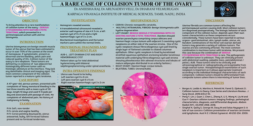
Objective: To bring attention to a rare manifestation of collision tumor of the ovary – SEROUS CYSTADENOMA WITH MATURE CYSTIC TERATOMA, which presented in a perimenopausal woman with uterine leiomyoma.
Introduction:
Uterine leiomyomas are benign smooth muscle tumor of the uterus that has been estimated to occur in over 70% of the women at the onset of menopause. If symptomatic, present as abnormal uterine bleeding, pelvic pain and reduced quality of life. Collison tumor of the ovary is rare neoplasm. These tumors are defined by co existence of two adjacent but histologically different tumor with no histologic admixture at the interface. They lack any identifiable clinical and imaging signs. The most common component of the collision tumor reported is a mature cystic teratoma.
Case presentation on A 47-year-old, parous woman, presented with complaints of heavy menstural bleeding in the last three months with a mean cycle of 30 days, length 10 days and used 5-6 pads per day and associated with passage of clots. No complaints of abdominal pain. No history of malignancy in family.
EXAMINATION:
P/A: Soft, non-tender
P/S: cervix and vagina healthy
P/V: Cervix pointing downwards, uterus anteverted, bulky, left forniceal fullness present and no forniceal tenderness.
INVESTIGATION:
- Hemogram revealed anemia.
- A transabdominal ultrasound revealed a anterior wall myoma of size 4 X 3 cm, a left ovarian cyst of 6 x 5 cm and a right haemorrhagic cyst of 3 x 2 cm.
- Biochemical investigations and the tumor markers were within the normal limits
Provisional diagnosis: AUB-L , LEFT OVARIAN CYST AND RIGHT HEMORRHAGIC CYST
Patient taken up for total abdominal hysterectomy with bilateral salphingoopherectomy under spinal anesthesia
Intra operative findings:
- Uterus was found to be bulky.
- Left ovarian cyst 6 x 6 cm
- Left para ovarian cyst 6 x 5 cm
- Right ovarian haemorrhagic cyst 3 x 2 cm
- Histopathology : CERVIX: Chronic nonspecific cervicitis.
- MULTIPLE INTRAMURAL FIBROID: Benign leiomyomata with hyaline degeneration
- LEFT OVARY: BENIGN SEROUS CYSTADENOMA WITH CO EXISTING MATURE CYSTIC TERATOMA. ( Section showed ovarian parenchyma comprising corpus albicans and haemorrhagic corpus luteum with adjacent 2 coexisting cystic neoplasms on either side of the ovarian stroma. One of the cystic neoplasm shows fibrocollagenous cyst wall lined by single layer of flattened cuboidal to ciliated columnar epithelium. Other cystic neoplasm is lined by keratinized stratified squamous epithelium enclosing lamellar keratinous material with underlying fibrocollagenous cyst wall stroma showing pilosebaceous skin adnexal structures and lobules of mature adipocytes distributed in an orderly fashion.
- RIGHT OVARY: haemorrhagic corpus luteum.
- BILATERAL TUBES: unremarkable.
Discussion: Uterine fibroids are common tumors affecting the perimenopausal women causing AUB. Most collision tumors are diagnosed postoperatively. The biologic behavior of each component of the collision tumor, depends upon their own tumor characteristics as these components occurs coincidentally These tumors have been reported in various organs- gastrointestinal, skin, lymph nodes, uterus, etc. Random combinations of the three-cell ovarian derived tumors may generate a variety of collision tumors. The ovaries are less commonly affected. The most common combination is mucinous tumor with teratoma. We present this case because the combination of SEROUS CYSTADENOMA WITH TERATOMA is rare. The pathogenesis of collision tumor is unknown. They can clinically present with abdominal swelling, palpable mass, pain(abdominal / pelvic), AUB. These tumors tend to be clinically and ultrasonographical or radiographically unrecognizable from single ovarian tumor. Histopathological examination of such neoplasm is important as it allows appropriate treatment according to the individual biological characteristics of each component. Collision tumors should be differentiated from composite tumors where there is intermixing of tumor lines.