1,830 posters, 67 videos, 75 audios, 29 topics, 22 sessions, 4,727 authors, 1,697 institutions
ePostersLive by SciGen Technologies S.A. All rights reserved.
RCOG World Congress 2023
June 12 - 14, 2023 | London, UK

EP.1533
CLASSICAL CASE OF PSEUDOMYXOMA PERITONEI SECONDARY TO MUCINOUS ADENOCARCINOMA OF THE OVARY.
Poster Presenter
Part of Topic
Gynae Oncology
CLASSICAL CASE OF PSEUDOMYXOMA PERITONEI SECONDARY TO MUCINOUS ADENOCARCINOMA OF THE OVARY.
MMH Dissanayake1, WMAPB Walisundara1, SSN Chandrasiri2, LGN Fernando1
1District General Hospital Matale, Sri Lanka
2De Soyza Hospital for Women, Colombo, Sri Lanka
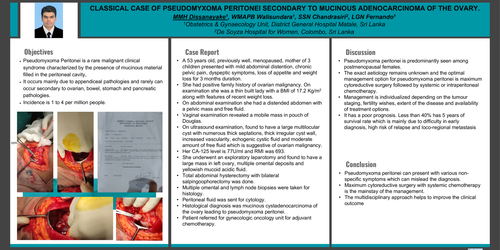
Objective: Pseudomyxoma Peritonei is a rare malignant clinical syndrome characterized by the presence of mucinous material filled in the peritoneal cavity, which occurs mainly due to appendiceal pathologies and rarely can occur secondary to ovarian, bowel, stomach and pancreatic pathologies. Incidence is 1 to 4 per million people.
Case report: A 53 years old, previously well, menopaused, mother of 3 children presented with mild abdominal distention, chronic pelvic pain, dyspeptic symptoms, loss of appetite and weight loss for 3 months duration. She had positive family history of ovarian malignancy. On examination she was a thin built lady with a BMI of 17.2 Kg/m2 along with features of recent weight loss. On abdominal examination she had a distended abdomen with a pelvic mass and free fluid. Vaginal examination revealed a mobile mass in pouch of Douglas. On ultrasound examination, found to have a large multilocular cyst with numerous thick septations, thick irregular cyst wall, increased vascularity, echogenic cystic fluid and moderate amount of free fluid which is suggestive of ovarian malignancy. Her CA-125 level is 77U/ml and RMI was 693. She underwent an exploratory laparotomy and found to have a large mass in left ovary, multiple omental deposits and yellowish mucoid acidic fluid. Total abdominal hysterectomy with bilateral salpingoophorectomy was done. Multiple omental and lymph node biopsies were taken for histology. Peritoneal fluid was sent for cytology. Histological diagnosis was mucinous cystadenocarcinoma of the ovary leading to pseudomyxoma peritonei. Patient referred for gynecologic oncology unit for adjuvant chemotherapy.
Discussion: Pseudomyxoma peritonei is predominantly seen among postmenopausal females. The exact aetiology remains unknown and the optimal management option for pseudomyxoma peritonei is maximum cytoreductive surgery followed by systemic or intraperitoneal chemotherapy. Management is individualized depending on the tumour staging, fertility wishes, extent of the disease and availability of treatment options. It has a poor prognosis. Less than 40% has 5 years of survival rate which is mainly due to difficulty in early diagnosis, high risk of relapse and loco-regional metastasis.
Conclusion: Pseudomyxoma peritonei can present with various non-specific symptoms which can mislead the diagnosis. Maximum cytoreductive surgery with systemic chemotherapy is the mainstay of the management. The multidisciplinary approach helps to improve the clinical outcome.