1,830 posters, 67 videos, 75 audios, 29 topics, 22 sessions, 4,727 authors, 1,697 institutions
ePostersLive by SciGen Technologies S.A. All rights reserved.
RCOG World Congress 2023
June 12 - 14, 2023 | London, UK

EP.1402
In-utero fetal bowel perforation: a case report.
Poster Presenter
Authors
Hagir Shalal, Miroslava Slavska, Catherine Stone, Amy Shacaluga
Affiliations
Part of Topic
Fetal Medicine
In-utero fetal bowel perforation: a case report
Dr Hagir Shalal, Ms. Miroslava Slavska, Ms Amy Shacaluga, Ms Catherine Stone
Aneurin Bevan University Health Board
Objective
Meconium peritonitis is a rare complication of in-utero fetal bowel perforation. With high index of clinical suspicion, prenatal ultrasound is a useful diagnostic tool. Congenital CMV may be an important etiological factor that needs to be considered.
Case report
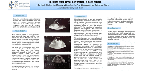
A 40- years old, G2 P1, 34 weeks, presented with abdominal pain and reduced fetal movements. She is fit and well had one vaginal delivery. Antenatal screening, dating and anomaly scan were normal. She and her partner are first cousins. She doesn’t smoke, has no allergies, has normal BMI. She was very uncomfortable with tense abdomen. Symphyseal fundal height was more than dates. CTG was normal. Ultrasound revealed a single fetus with normal doppler, head and femur circumferences. There was polyhydramnios with amniotic fluid index of 27 cm. Abdominal circumference was 509 mm, showing fetal ascites and pericardial effusion. Intrauterine fetal infection was suspected, and TORCH screen was carried out. Emergency cesarean section was done for maternal discomfort. The baby was admitted to NICU.
Discussion
Meconium peritonitis is rare and occurs in approximately 1/35,000 live birth (1-3). It’s a sterile chemical peritonitis resulting from small bowel perforation In utero, usually presenting during or after the second trimester (4). The association of meconium peritonitis with congenital CMV, rubella and parvovirus B-19 infections is documented (5-7). In this case both baby and mother were seropositive for CMV, and the mother was also positive for rubella and parvovirus B-19. The peritonitis results from intestinal fluid and meconium extruding into the peritoneal cavity through a perforation in the fetal intestine. The inflamed bowel loops conceal the inflammation leading to formation of meconium pseudocyst and dystrophic intraperitoneal calcifications (8). Fetal ascites was reported as the commonest ultrasound finding, pseudocyst in 28.5%, and polyhydramnios in 50%. It was reported fetal ascites and either calcifications or dilated bowel loops are the minimal criteria for perinatal diagnosis of meconium peritonitis (8-10). In this case fetal ascites and polyhydramnios were clearly seen in the perinatal ultrasound. Neonatal images revealed ascites and curvilinear peritoneal calcifications.The baby had laparotomy and jejunostomy. Intra-operatively, there were ascites, meconium pseudocyst, jejunal volvulus, and perforation which are keeping with the perinatal and neonatal radiological findings.
Conclusion
In-utero bowel perforation with meconium peritonitis is rare. Fetal ascites and intra-abdominal calcifications are the commonest ultrasound findings. CMV is an important etiological consideration, further studies are needed in this field
References
1. Olsen MM, Luck SR, Lloyd-Still J, Raffensperger JG. The spectrum of meconium disease in infancy. J Pediatr Surg 1982;17:479–81.
2. Pan EY, Chen LY, Yang JZ, Lee Z, Wang ZZ. Radiographic diagnosis of meconium peritonitis. A report of 200 cases including six fetal cases. Pediatr Radiol 1983;13:199–205.
3. Payne RM, Nielsen AM. Meconium peritonitis. Am Surg 1962;28:224–31.
4/ Dἃhnert W. Radiology review manual.6th ed. Philadelphia: Lippincott Williams & Wilkins; 2007; p. 856
5/ Reynolds E, Douglass B, Bleacher J. Meconium peritonitis. J Perinatol 2000;20:193–5.
6/ Radner M, Vergesslich KA, Weninger M, Eilenberger M, Ponhold W, Pollak A. Meconium peritonitis: a new finding in rubella syndrome. J Clin Ultrasound 1993;21:346–9.
7/ Schild RL, Plath H, Thomas P, Schulte-Wissermann H, Eis-Hubinger AM, Hansmann M. Fetal parvovirus B19 infection and meconium peritonitis. Fetal Diagn Ther 1998;13:15–8.
8/ Alessandro P et al, 2016. Neonatal gastrointestinal involvement and congenital cytomegalovirus. La Paediatria medica e Chirurgica, vol. 38, p. 134.
9/ Chan KL, Tang MH, Tse HY, Tang RY, Tam PK. Meconium peritonitis: prenatal diagnosis, postnatal management and outcome. Prenat Diagn. 2005;25:676–82.
10/ Dirkes K, Crombleholme TM, Craigo SD, Latchaw LA, Jacir NN, Harris BH, D’Alton ME. The natural history of meconium peritonitis diagnosed in utero. J Pediatr Surg 1995; 30:979–82