1,830 posters, 67 videos, 75 audios, 29 topics, 22 sessions, 4,727 authors, 1,697 institutions
ePostersLive by SciGen Technologies S.A. All rights reserved.
RCOG World Congress 2023
June 12 - 14, 2023 | London, UK

EP.1391
Prolapsed pedunculated fibroid, suspected as cervical malignancy on imaging.
Poster Presenter
Authors
Affiliations
Part of Topic
Benign Gynaecology
Prolapsed pedunculated fibroid, suspected as cervical malignancy on imaging.
Dr. Aisha Nisar and Miss.Rakhee Saxena
Nottingham University Hospitals NHS trust.
To emphasize the importance of thorough clinical examination while considering imaging as a useful adjuvant to reach clinical diagnosis. To highlight one of the rare presentation of continuous abnormal vaginal bleeding as prolapsed pedunculated submucous fibroid.
32 years old lady, para3 with all vaginal deliveries, presented in ED with four-months h/o heavy vaginal bleeding. She had mild lower abdominal pain and feeling of dizziness and lethargy. She tried tranexamic acid and nor-ethisterone with the GP that did not help and was currently awaiting a scan.Eventually her bleeding became heavy and she was changing pads every hour. She was regular with her smears and last smear was normal.
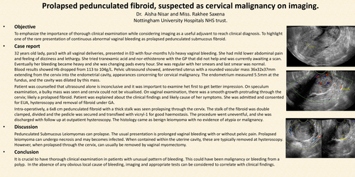
Blood results showed Hb dropped from 113 to 104g/L. Pelvic ultrasound showed, anteverted uterus with a rounded vascular mass 36x32x37mm extending from the cervix into the endometrial cavity, appearances concerning for cervical malignancy. The endometrium measured 5.5mm at the fundus, and the cavity was dilated by this mass.
Patient was counselled that ultrasound alone is inconclusive and it was important to examine het first to get better impression. On speculum examination, a bulky mass was seen and cervix could not be visualised. On vaginal examination, there was a smooth growth protruding through the cervix; likely a prolapsed fibroid. Patient was explained about the clinical findings and likely cause of her symptoms. She was admitted and consented for EUA, hysteroscopy and removal of fibroid under GA.
Intra-operatively, a 6x8 cm pedunculated fibroid with a thick stalk was seen prolapsing through the cervix. The stalk of the fibroid was double clamped, divided and the pedicle was secured and transfixed with vicryl-1 for good haemostasis. The procedure went uneventful, and she was discharged with follow up at outpatient hysteroscopy. The histology came as benign leiomyoma with no evidence of atypia or malignancy.
Pedunculated Submucous Leiomyomas can prolapse. The usual presentation is prolonged vaginal bleeding with or without pelvic pain.Prolapsed leiomyoma can undergo necrosis and may becomes infected. When contained within the uterine cavity, these are typically removed at hysteroscopy. However, when prolapsed through the cervix, can usually be removed by vaginal myomectomy.
It is crucial to have thorough clinical examination in patients with unusual pattern of bleeding. This could have been malignancy or bleeding from a polyp. In the absence of any obvious local cause of bleeding, imaging and appropriate tests can be considered to correlate with clinical findings.