1,830 posters, 67 videos, 75 audios, 29 topics, 22 sessions, 4,727 authors, 1,697 institutions
ePostersLive by SciGen Technologies S.A. All rights reserved.
RCOG World Congress 2023
June 12 - 14, 2023 | London, UK

EP.1139
Incidental Finding of Endometriosis at Caesarean Section
Poster Presenter
Authors
Affiliations
Part of Topic
High Risk Pregnancy
Incidental Finding of Endometriosis at Caesarean Section
Jordan-Brown D. Romeo-Bassey R, Ramdass J
OBJECTIVE
Endometriosis is the growth of endometrial tissue outside of the uterus. It is known to cause subfertility, but women who do conceive, their pregnancies can be complicated. The risk of miscarriage, placenta praevia, fetal growth restriction, pre-eclampsia, preterm birth (PTB), premature rupture of membranes and spontaneous haemoperitoneum in pregnancy are increased. This case report aims to highlight the complications of endometriosis in pregnancy.
CASE REPORT
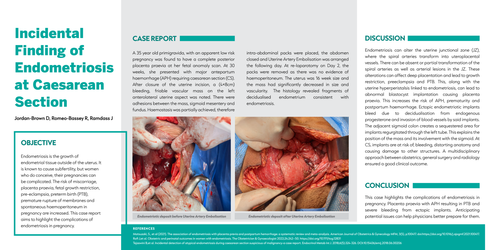
A 35 year old primigravida, with an apparent low risk pregnancy was found to have a complete posterior placenta praevia at her fetal anomaly scan. At 30 weeks, she presented with major antepartum haemorrhage (APH) requiring caesarean section (CS). After closure of the uterine incision, a (4×8cm) bleeding, friable vascular mass on the left anterolateral uterine aspect was noted. There were adhesions between the mass, sigmoid mesentery and fundus. Haemostasis was partially achieved, therefore intra-abdominal packs were placed, the abdomen closed and Uterine Artery Embolisation was arranged the following day. At re-laparotomy on Day 2, the packs were removed as there was no evidence of haemoperitoneum. The uterus was 16 week size and the mass had significantly decreased in size and vascularity. The histology revealed fragments of decidualised endometrium consistent with endometriosis.
DISCUSSION
Endometriosis can alter the uterine junctional zone (JZ), where the spiral arteries transform into uteroplacental vessels. There can be absent or partial transformation of the spiral arteries as well as arterial lesions in the JZ. These alterations can affect deep placentation and lead to growth restriction, preeclampsia and PTB. This, along with the uterine hyperperistalsis linked to endometriosis, can lead to abnormal blastocyst implantation causing placenta praevia. This increases the risk of APH, prematurity and postpartum haemorrhage. Ectopic endometriotic implants bleed due to decidualisation from endogenous progesterone and invasion of blood vessels by said implants. The adjacent sigmoid colon creates a sequestered area for implants regurgitated through the left tube. This explains the position of the mass and its involvement with the sigmoid. At CS, implants are at risk of, bleeding, distorting anatomy and causing damage to other structures. A multidisciplinary approach between obstetrics, general surgery and radiology ensured a good clinical outcome.
CONCLUSION
This case highlights the complications of endometriosis in pregnancy. Placenta praevia with APH resulting in PTB and severe bleeding from ectopic implants. Anticipating potential issues can help physicians better prepare for them.