1,830 posters, 67 videos, 75 audios, 29 topics, 22 sessions, 4,727 authors, 1,697 institutions
ePostersLive by SciGen Technologies S.A. All rights reserved.
RCOG World Congress 2023
June 12 - 14, 2023 | London, UK

EP.1138
POSTERIOR REVERSIBLE ENCEPHALOPATHY SYNDROME ASSOCIATED WITH ECLAMPSIA
Poster Presenter
Part of Topic
Maternal Medicine
POSTERIOR REVERSIBLE ENCEPHALOPATHY SYNDROME ASSOCIATED WITH ECLAMPSIA
Kamalathasan P, Mahanama A, Jayarathna RASH, Jayantha N, Amaradivakara PW -Teaching Hospital Mahamodara, Sri Lanka
Introduction: Posterior reversible encephalopathy syndrome (PRES) is a clinical and radiological diagnosis of a rare condition. It is clinically presented with headache, altered consciousness, visual disturbances, seizures and nausea and is radiologically characterized by vasogenic oedema in the posterior circulation of the brain1. In addition, a patient may have hemianopia, paresis, dysphasia and other focal neurological deficits. Toxaemia in pregnancy, hypertension, autoimmune disease, renal failure and cytotoxic and immunosuppressive medications are the common precipitating factor for this condition. PRES is a reversible condition if diagnosed early. The pathophysiology of PRES is unknown but attributed to brain oedema secondary to endothelial dysfunction and failure in cerebral blood pressure autoregulation in the posterior circulation of the brain2.
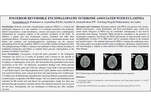
Case Presentation: Unconscious 18-year-old primi presented to the Emergency Treatment Unit (ETU) at 32 weeks of gestation with a history of one episode of convulsion. She didn’t have any regular antenatal follow-ups, and there was no history of epilepsy or hypertension in her past. She had another four generalized tonic-clonic convulsions at the ETU. On admission, examination findings were blood pressure (BP)- 200/110 mmHg, pulse rate-110b/min, Glasgow Coma Scale-8 and urine albumin 4+ and Blood reports were Haemoglobulin level-12g/dl, platelet count-360,000/mm3, Uric acid level-584mic mol/l, normal limit liver and renal function tests. Furthermore, CT finding was ill-defined hypo densified areas involving bilateral symmetrical parito- occipital regions compatible with PRES changes. She was treated with Hydralazine and Labetalol for BP control and with Mgso4 for neuroprotection. Single live fetus was delivered through emergency caesarian section. Post-operative intensive care was given for five days. Subsequently, she was discharged on following day after complete recovery.
Discussion and Conclusion: Eclamptic patients with PRES may present with seizures, altered consciousness, visual disturbances and focal neurological signs, which may mimic stroke. Diagnosis of PRES may be overlooked. Subsequently, it may lead to irreversible brain damage. Therefore, PRES should be considered in the presence of neurological symptoms, risk factors for PRES and absence of other possible causes of encephalopathy. In brain CT or MRI, there will be vasogenic oedema in the occipital and parietal regions. The standard treatment for PRES is treating the underlying cause. This index patient was treated with BP control and delivery. Meticulous clinical analysis and neuroimaging is helpful in early detection of PRES and prevention of permanent brain damage.