1,830 posters, 67 videos, 75 audios, 29 topics, 22 sessions, 4,727 authors, 1,697 institutions
ePostersLive by SciGen Technologies S.A. All rights reserved.
RCOG World Congress 2023
June 12 - 14, 2023 | London, UK

EP.1125
Non Puerperal Acute Uterine Inversion : A Diagnostic Dilemma and Surgical Challenge
Poster Presenter
Authors
Affiliations
Part of Topic
Benign Gynaecology
Audio
Non Puerperal Acute Uterine Inversion: A Diagnostic Dilemma and Surgical Challenge
Noura Aldaham, Ayesha Arif, Bilquisunnisa Begum
King Saud Medical City, Riyadh, Saudi Arabia
Introduction:
Non Puerperal Uterine Inversion (NPUI), a rare clinical condition, has an unknown incidence owing to scarcity of available literature. We describe an unusual case of acute uterine inversion due to submucous myoma in a postmenopausal lady, managed successfully with prompt diagnosis and timely surgical intervention.
Objective :
We aim to highlight the atypical presentation, diagnostic limitations and preferred surgical approach for this rare clinical challenge.
Case Report :
A 53 year old postmenopausal lady with history of five uncomplicated vaginal births presented to the gynecological emergency department with sudden onset of abdominal pain, mild vaginal bleeding and protruding vulval mass for two days. Known case of diabetes, hypertension and end stage renal disease; her past gynecological history was unremarkable except for mild suprapubic tenderness. Vulval examination revealed a large fungating mass protruding out of vaginal introitus with a thick pedicle and bloody discharge; the cervical os couldn’t be identified. Uterus wasn’t palpable on rectal examination.
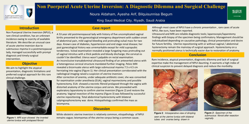
An inconclusive transabdominal ultrasound finding of an anteverted uterus with a heterogenous cervical structure mandated further imaging. Pelvic MRI revealed a bicornuate uterus with large submucosal myoma 7.5 x 4.5 cm herniating into vagina (Figure 1). Her clinical presentation corroborated with the radiological imaging raised a suspicion of uterine inversion.
After correction of anemia, under adequate antibiotic cover, she was consented for examination under anesthesia (EUA), vaginal myomectomy and possible hysterectomy. EUA showed a necrotic fibroid prolapsed through the vagina with distorted anatomy of the uterine corpus and cervix. We proceeded with exploratory laparotomy to confirm uterine inversion (Figure 2) and restore the anatomy. Vaginal resection of the myoma (Figure 3) was followed by successful uterine repositioning. Total abdominal hysterectomy with bilateral salpingoophorectomy was done. Histopathology confirmed the mass as leiomyoma.
Discussion :
While obstetric uterine inversion is relatively common, etiopathology of NPUI remains vague; leiomyomas of the uterine corpus being a common cause.
Although most cases of NPUI have a chronic presentation , rare cases of acute NPUI, like ours, have been reported.
Ultrasound and MRI are reliable diagnostic tools; laparoscopic/laparotomy findings with biopsy of the specimen being confirmatory. Management should be individualized depending on causative pathology, clinical presentation and desire for future fertility. Uterine repositioning with or without vaginal / abdominal hysterectomy remain the mainstay of surgical approach. Hysterectomy on a normally positioned uterus is technically easier due to restoration of anatomy.
Conclusion :
Rare incidence, atypical presentation, diagnostic dilemma and lack of surgical expertise make the management of NPUI daunting. It warrants a high index of clinical suspicion to prevent delayed diagnosis and reduce the morbidity.