1,830 posters, 67 videos, 75 audios, 29 topics, 22 sessions, 4,727 authors, 1,697 institutions
ePostersLive by SciGen Technologies S.A. All rights reserved.
RCOG World Congress 2023
June 12 - 14, 2023 | London, UK

EP.1085
Surgical management of symptomatic maternal hydronephrosis: is it worth it?
Poster Presenter
Authors
Affiliations
Part of Topic
High Risk Pregnancy
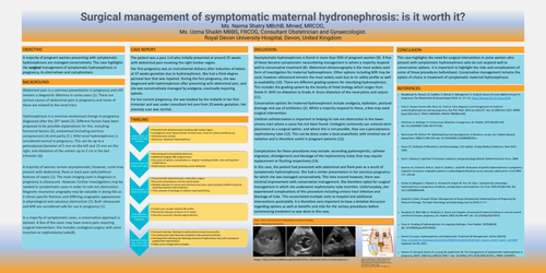
SURGICAL MANAGEMENT OF SYMPTOMATIC MATERNAL HYDRONEPHROSIS: IS IT WORTH IT?
Ms. Naima Shatry MBchB, Mmed, MRCOG,
Ms. Uzma Shaikh MBBS, FRCOG, Consultant Obstetrician and Gynaecologist. Royal Devon University Hospital, Devon, United Kingdom
OBJECTIVE:
A majority of pregnant women presenting with symptomatic hydronephrosis are managed conservatively. This case highlights the surgical management of symptomatic hydronephrosis in pregnancy, its alternatives and complications.
BACKGROUND:
Abdominal pain is a common presentation in pregnancy and still remains a diagnostic dilemma in some cases (1). There are various causes of abdominal pain in pregnancy and some of these are related to the renal tract. Hydronephrosis is a common anatomical change in pregnancy diagnosed after the 20th week (2). Different factors have been proposed to be possible explanations for this, including: hormonal factors (3), anatomical (including extrinsic compression) (4) and parity (5 ). Mild renal hydronephrosis is considered normal in pregnancy. This can be up to a pelvicalyceal diameter of 5 mm on the left and 15 mm on the right, and dilatation of the ureters up to 2 cm in the last trimester (6). A majority of women remain asymptomatic; however, some may present with abdominal, flank or back pain with/without features of sepsis (1). The main imaging used in diagnosis in pregnancy is ultrasound. However, further investigations may be needed in symptomatic cases in order to rule out obstruction. Magnetic resonance urography may be valuable in doing this as it shows specific features and differing urographic appearances in physiological and calculous obstruction (7). Both ultrasound and MRI are considered safe for use in pregnancy (1). In a majority of symptomatic cases, a conservative approach is advised. A few of the cases may have severe pain requiring surgical intervention: this includes urological surgery with stent insertion or nephrostomy tube(8).
CASE REPORT:
The patient was a para 1+0 who initially presented at around 25 weeks with abdominal pain involving the right lumbar region. Her first pregnancy was an instrumental delivery after induction of labour at 37 weeks gestation due to hydronephrosis. She had a third-degree perineal tear that was repaired. During the first pregnancy, she was diagnosed with hydronephrosis after presenting with abdominal pain, and she was conservatively managed by analgesia, eventually requiring opioids. For her current pregnancy, she was booked by the midwife in her first trimester and was under consultant led care from 20 weeks gestation. Her anomaly scan was normal.
TIMELINE:
Initial contact: 25 weeks and 3 days Gestation
• Presented with abdominal pain involving right lumbar region.
• Investigations done: Renal and liver function tests, Urine for culture and Renal scan.
• All bloods were normal.
• Renal scan- Moderate Hydronephrosis
Second contact: 26 weeks and 6 days
• Worsening pain necessitating admission.
• Additional imaging: MRI Urogram done.
• Discussion of options: Conservative vs. Surgical- Including benefits, risks and long term complications.
• Nephrostomy tube inserted during current admission.
Third contact: 27 weeks and 6 days
• Presented with abdominal pain a week after surgery.
• Recurrent presentations over the next 8 weeks.
• Multiple episodes of urinary tract infections that were culture positive (coliform bacteria) requiring treatment with antibiotics.
• Three episodes of blockage of catheter requiring flushing.
Antenatal Clinic contact:36 weeks Gestation
• Growth scan: Growth noted at 9th centile
• Planned for induction of labour at 37 weeks.
• Induction successful- Normal vaginal delivery.
Postnatal contact: Day 1 post natal and at two weeks.
• First post-natal day: Attempt at nephrostomy removal unsuccessful.
• Two weeks post-natal: Removal completed under general anesthetic.
• Discharged the following day following removal of nephrostomy tube with subsequent symptomatic improvement.
• Follow up by Urology team arranged.
DISCUSSION:
Asymptomatic hydronephrosis is found in more than 90% of pregnant women (9). A few of these become symptomatic necessitating management in whom a majority respond well to conservative treatment (8). Abdominal ultrasonography is the most widely used form of investigation for maternal hydronephrosis. Other options including MRI may be used, however ultrasound remains the most widely used due to its safety profile as well as availability (10). There are different grading systems for classifying hydronephrosis. This includes the grading system by the Society of Fetal Urology which ranges from Grade 0- With no dilatation to Grade 4- Gross dilatation of the renal pelvis and calyces (11). Conservative options for maternal hydronephrosis include analgesia, hydration, postural drainage and use of antibiotics (2). Whilst a majority respond to these, a few may need surgical intervention. Urethral catheterisation is important in helping to rule out obstruction in the lower urinary tract where a cause has not been found. Urologists commonly use ureteral stent placement as a surgical option, and where this is not possible, they use a percutaneous nephrostomy tube (12). This can be done under a local anaesthetic with minimal use of radiation and is therefore useful in pregnant patients (12). Complications for these procedures may include: ascending pyelonephritis, catheter migration, dislodgement and blockage of the nephrostomy tubes that may require replacement or flushing respectively (13). In this case, the patient had presented with abdominal and flank pain as a result of symptomatic hydronephrosis. She had a similar presentation in her previous pregnancy for which she was managed conservatively. This time around however, there was minimal improvement with conservative management. She therefore opted for surgical management in which she underwent nephrostomy tube insertion. Unfortunately, she experienced complications of this procedure including urinary tract infection and blockage of tube. This necessitated multiple visits to hospital and additional interventions postnatally. It is therefore very important to have a detailed discussion regarding options as well as benefits and risks for the various procedures before commencing treatment as was done in this case.
CONCLUSION:
This case highlights the need for surgical intervention in some women who present with symptomatic hydronephrosis who do not respond well to conservative options. It is important to highlight the risks and complications of some of these procedures beforehand. Conservative management remains the option of choice in treatment of symptomatic maternal hydronephrosis.
REFERENCES:
1. Woodhead, N, Nkwam, O, Caddick, V, Morad, S, Mylvaganam, S. Surgical causes of acute abdominal pain in pregnancy. The Obstetrician & Gynaecologist 2019; 21: 27– 35. https://doi.org/10.1111/tog.12536
2. Ciciu E, Paṣatu-Cornea AM, Petcu LC, Tuţă LA. Early diagnosis and management of maternal ureterohydronephrosis during pregnancy. Exp Ther Med. 2022 Jan;23(1):27. doi: 10.3892/etm.2021.10949. Epub 2021 Nov 5. PMID: 34824635; PMCID: PMC8611492.
3. Schulman A, Herlinger H. Urinary tract dilatation in pregnancy. Br J Radiol. 1975;48:638–645. doi: 10.1259/0007-1285-48-572-638.
4. Rasmussen PE, Nielsen FR. Hydronephrosis during pregnancy: A literature survey.Eur J Obstet GynecolReprod Biol. 1988;27:249–259. doi: 10.1016/0028-2243(88)90130-x.
5. Dawn CS. Textbook of Obestetrics and Neonatology. 11th edition. Pratap Medical Publishers, New Delhi, 1995.
6. Sarris I, Bewley S, Agnihotri S.Training in obsterics and gynaecology.Oxford: Oxford University Press; 2009
7. Spencer JA, Chahal R, Kelly A, Taylor K, Eardley I, Lloyd SN. Evaluation of painful hydronephrosis in pregnancy: magnetic resonance urographic patterns in physiological dilatation versus calculous obstruction J Urol 2004; 171: 256-60.
8. Puškar D, Balagović I, Filipović A, Knezović N, Kopjar M, Huis M, Gilja I. Symptomatic physiologic hydronephrosis in pregnancy: Incidence, complications and treatment. Eur Urol. 2001;39:260–263. doi: 10.1159/000052449.
9. Sudanshu Citale, Vinayak Chitale, Management of Acute Symptomatic Hydronephrosis of Pregnancy by Postural Drainage, The Open Neurology and Nephrology Journal 2010; 3: 4-7.
10. Karabulut N, Baki Yaǧci A, Karabulut A. Renal vein Doppler ultrasound of maternal kidneys in normal second and third trimester pregnancy. Br J Radiol. 2003;76:444–447. doi: 10.1259/bjr/81976752.
11. Onen A. Grading of hydronephrosis: An ongoing challenge. Front Pediatr. 2020;8(458) doi: 10.3389/fped.2020.00458.
12. Dennis G Lusaya, Hydronephrosis and Hydroureter Treatment & Management. Article 436259, https://emedicine.medscape.com/article/436259 treatment?icd=login_success_email_match_norm#d8Updated: Oct 03, 2022.
13. Fainaru O, Almog B, Gamzu R, Lessing JB, Kupferminc M. The management of symptomatic hydronephrosis in pregnancy. BJOG. 2002 Dec;109(12):1385-7. doi: 10.1046/j.1471-0528.2002.01545.x. PMID: 12504975.