1,830 posters, 67 videos, 75 audios, 29 topics, 22 sessions, 4,727 authors, 1,697 institutions
ePostersLive by SciGen Technologies S.A. All rights reserved.
RCOG World Congress 2023
June 12 - 14, 2023 | London, UK

EP.1071
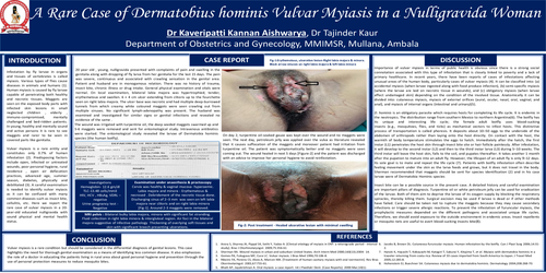
A rare case of Dermatobius hominis Vulvar Myiasis in a nulligravida woman
Part of Topic
Benign Gynaecology
Introduction : Infestation by fly larvae in organs and tissues of vertebrates is called myiasis. Various types of flies cause diseases in animals and humans (1). Human myiasis is caused by fly larvae capable of penetrating both healthy and necrotic tissues. Maggots are seen on the exposed body parts with infected skin lesions in small neglected children , elderly , immuno-compromised, mentally challenged and bed-ridden patients. In young, healthy and mentally sound and active persons it is rare to see maggots and rarer to be seen in covered parts like genitalia.
Vulvar myiasis is a rare entity and constitutes only 0.7% of human infestation (2). Predisposing factors include open, infected or untreated wounds, poor personal hygiene, rural residence , open air defecation practices, advanced age, summer season, diabetes, physically and debilitated (3). A careful examination is needed to identify vulvar myiasis as it can be confused with many common diseases such as insect bite, cellulitis, etc. Here we report the rare case of vulvar myiasis in a 20-year-old educated nulligravida with sound physical and mental health status.
case report :
20 year old , young, nulligravida presented with complaints of pain and swelling in the genitalia along with dropping of fly larva from her genitalia for the last 15 days. The pain was severe, continuous and associated with crawling sensation in the genital area. Patient and husband are in monogamous relation. There was no history of trauma, insect bite, chronic illness or drug intake. General physical examination and vitals were normal. On local examination, bilateral labia majora was hypertrophied, tender, erythematous and swollen. 6 × 4 cm ulcer extending from clitoris up to the fourchette seen on right labia majora. The ulcer base was necrotic and had multiple deep-burrowed tunnels from which creamy white coloured maggots were seen crawling out from multiple sinuses. No significant lymph-adenopathy was present. The husband was examined and investigated for similar signs or genital infections and revealed no evidence of the same .
The wound was irrigated with turpentine oil, the deep seeded maggots swarmed up and 5-6 maggots were removed and sent for entomological study. Intravenous antibiotics were started. The entomological study revealed the larvae of Dermatobia hominis species (the human botfly).
Examination under anaesthesia & proctoscopy: Cervix was healthy & vaginal mucosa- hyperaemic. Labia majora and minora - Erythematous & necrosed . Debridement of the necrotic tissue done. Discharging sinus of 2–3 mm was seen on left labia majora near clitoris and on right labia minora [Fig-1]. Around 2-3 maggots were removed.
MRI pelvis : Bilateral bulky labia majora, minora with significant fat stranding.Fluid collection in right labia minora & intergluteal region. Air foci in the bilateral majora suggestive of infective pathology Thickening of overlying soft tissues and skin with significant breech presenting ulcerations.
On day 2, turpentine oil-soaked gauze was kept over the wound and no maggots were seen. The next day, petroleum jelly was applied over the vulva as literature revealed that it causes suffocation of the maggots and moreover patient had irritation from turpentine oil. The patient was symptomatically better and no maggots were seen coming out. The wound healed in next 5 days [Figure-2] and the patient was discharged with an advice to improve her personal hygiene to avoid reinfestation.
Discussion : Importance of vulvar myiasis in terms of public health is obvious since there is a strong social connotation associated with this type of infestation that is closely linked to poverty and a lack of primary healthcare. In recent years, there have been reports of cases of infestations affecting unusual areas of the human body, particularly cases of vulvar myiasis (4). It can be classified into: (a) accidental myiasis (when larvae ingested along with food produce infection), (b) semi-specific myiasis (where the larvae are laid on necrotic tissue in wounds), and (c) obligatory myiasis (when larvae affect undamaged skin) depending on the condition of the involved tissue. Anatomically it can be divided into: cutaneous myiasis, myiasis of external orifices (aural, ocular, nasal, oral, vaginal, and anal), and myiasis of internal organs (intestinal and urinary)(5).
D. hominis is an obligatory parasite which requires hosts for completing its life cycle. It is endemic in the neotropics. The distribution range from southern Mexico to northern Argentina(6). The botfly has its unique and interesting life cycle; the female adult botfly uses blood-sucking insects (e.g., mosquitoes, flies, and ticks) as mechanical vectors to infest hosts. This distinctive process of transportation is called phoresis. It deposits about 10–50 eggs to the underside of the abdomen of arthropods rather than laying onto the host directly. On contact with the host, the warmth of body temperature stimulates the eggs to hatch. Immediately after the hatch, the first instar (L1) penetrates the host skin through insect bite site or hair follicle painlessly. After infestation, it will develop to the second instar (L2) and then to the third instar larva (L3) during 5–10 weeks. The third instar will leave the host skin, falls to the soil, and pupates thereafter. It takes another 1 month after the pupation to mature into an adult fly. However, the lifespan of an adult fly is only 9–12 days. Its sole goal is to mate and repeat the life cycle (7). Patients with botfly infestation often describe feeling movement under the skin as the larva feeds and grows, but it does not travel in the body. Sherman recommended that maggots should be sent for species identification (2) and in his case larvae were of Dermatobia Hominis species
Insect bite can be a possible source in the present case. A detailed history and careful examination are important pillars of diagnosis. Turpentine oil or white petroleum jelly can be used for eradication of the larvae. Both of them aim to deprive the larvae of its oxygen supply by blocking the respiratory spiracles, thereby killing them. Surgical excision may be used if larvae is dead or if other methods have failed. Care should be taken not to rupture the maggots because they may cause secondary infections or trigger severe allergic reactions. To prevent the infestation of furuncular myiasis, the prophylactic measures depended on the different pathogens and associated unique life cycles. Therefore, we should avoid exposure to the outside environment in endemic areas. Insect repellents or mosquito nets are useful to avert blood-sucking insects bite(8).
Conclusion :Vulvar myiasis is a rare condition but should be considered in the differential diagnosis of genital lesions. This case highlights the need for thorough genital examination as a means of identifying less common disease. It also emphasizes the role of a doctor in educating the patients living in rural area about good personal hygiene and prevention through the use of personal protection measures to reduce mosquito bites.
References :