1,830 posters, 67 videos, 75 audios, 29 topics, 22 sessions, 4,727 authors, 1,697 institutions
ePostersLive by SciGen Technologies S.A. All rights reserved.
RCOG World Congress 2023
June 12 - 14, 2023 | London, UK

EP.1070
Intra-uterine death and ‘metabolic mayhem’: a case report
Poster Presenter
Authors
Affiliations
Part of Topic
Maternal Medicine
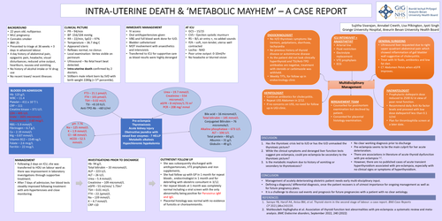
INTRA-UTERINE DEATH & ‘METABOLIC MAYHEM’ – A CASE REPORT
BACKGROUND
•22 years old, nulliparous
•MLC pregnancy
•Cannabis smoker
•BMI 25
•Presented to triage at 36 weeks + 3 days in advanced labour.
•4 day history of abdominal pain, epigastric pain, headache, visual disturbances, reduced urine output, heartburn, nausea and vomiting
•No history of alcohol intake or IV drug use
•No recent travel/ recent illnesses
CLINICAL PICTURE
•PR – 94/min
•BP -156/104 mmHg
•RR – 22/min, SpO2 – 97%
•Temperature – 36.7 deg C
•Appeared icteric
•Reflexes normal, no clonus
•Local examination- Vertex visible on perineum
•Ultrasound – No fetal heart beat detected.
•Intra-uterine death confirmed by 2 doctors.
•Stillborn male infant born by SVD with birth weight 2280g (< 5th percentile).
IMMEDIATE MANAGEMENT
•IV access
•Oral antihypertensives given
•VBG and full blood work done for IUD.
•Bladder catheterised
•MDT involvement with anaesthetics and intensivists
•Transferred to ICU for supportive care as blood results were highly deranged
AT ICU
•GCS – 15/15
•CVS – Ejection systolic murmur+
•RS – B/L air entry +, no added sounds
•P/A – soft, non-tender, uterus well contracted
•Lochia - NAD
•Poor urine output: 0-10ml/hr
•No headache or blurred vision
MANAGEMENT
•Following 2 days on ICU, she was transferred to HDU on labour ward as there was improvement in laboratory investigations through supportive treatment.
•After 7 days of admission, her blood tests steadily improved following treatment with anti-hypertensives and close monitoring.
OUTPATIENT FOLLOW UP
•She was subsequently discharged with antihypertensives, VTE prophylaxis and iron supplements.
•She had follow up with GP in 1 month for repeat bloods , endocrinologist in 1 month and for debriefing with obstetric consultant in 3/12.
•Her repeat bloods at 1 month was completely normal including a viral screen with the only abnormality being positive for Parvovirus IgM and IgG.
•Placental histology was normal with no evidence of funisitis or chorioamnionitis.
Discussion:
1.Has the thyrotoxic crisis led to IUD or has the IUD unmasked the thyrotoxic picture?
2.While the clinical symptoms and deranged liver function tests suggest pre-eclampsia, could pre-eclampsia be secondary to the thyrotoxic picture?
3.Is the metabolic mayhem due to history of vomiting or secondary to thyrotoxicosis?
•No clear working diagnosis prior to discharge
•Pre-eclampsia seems to be the main culprit for her acute deterioration.
•There are associations in literature of acute thyroid dysfunction with pre-eclampsia 1,2.
•However, there are no published cases of acute transient hyperthyroidism associated with pre-eclampsia, especially with no clinical signs or symptoms of hyperthyroidism.
Conclusion
•Management of acutely deteriorating obstetric patient needs early multi-disciplinary input.
•Defining a diagnosis/ differential diagnosis, once the patient recovers is of utmost importance for ongoing management as well as for future pregnancy plans.
•It is a challenge to discuss sad events and prognosis for future pregnancies with a patient with no clear aetiology.