1,830 posters, 67 videos, 75 audios, 29 topics, 22 sessions, 4,727 authors, 1,697 institutions
ePostersLive by SciGen Technologies S.A. All rights reserved.
RCOG World Congress 2023
June 12 - 14, 2023 | London, UK

EP.1069
An intraoperative surprise – a case report of a Retroperitoneal Mullerian Cyst
Poster Presenter
Authors
Philippa Dann, Phalguni Kotabagi, Kimberley Raynsford, Sunil Doshi
Affiliations
Part of Topic
Benign Gynaecology
An intra-operative surprise – a case report of Retroperitoneal Mullerian Cyst
Objective:
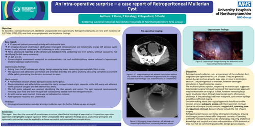
To describe a retroperitoneal cyst, identified unexpectedly intra-operatively. Retroperitoneal cysts are rare with incidence of 1/5750 to 1/250,000; one third are asymptomatic and incidental findings.
Case report:
à CT imaging showed small bowel obstruction (managed conservatively) and incidentally a large left adnexal cystic lesion; simple, without septations, wall thickening or solid components.
à Pelvic ultrasound reported a left adnexal cyst (82x80x75mm), containing low-level echoes, without vascularity; not identifying the left ovary separately.
à CA 125 was 11.
Laparoscopic findings:
Open surgery:
Histology:
Discussion:
Retroperitoneal mullerian cysts are remanent of the mullerian duct, diagnosed pre-operatively in 25% of cases. They are generally serous, benign and can grow to large sizes or recur in incomplete excision. The pathogenesis is unknown, however oestrogen stimulation is hypothesised as a cause.
The multidisciplinary opinion, suspecting an ovarian cyst, suggested laparoscopic surgical removal. Success of this laparoscopic approach may be dependent on surgical skillset, however removing large cystic structures intact, through laparoscopic ports often poses challenge. If the pathology had been malignant, cyst content spillage would have affected staging.
Decision making about the surgical approach should ensure the incision achieves adequate access and intact specimen retrieval. Operative technique should consider: surgical skill, operating time and specimen retrieval. Consent should include the risk of open surgery.
Retroperitoneal masses can mimic other pelvic structures, proving that imaging cannot always offer diagnostic certainty. Operating within the retroperitoneum can be challenging, requiring anatomical knowledge and surgical precision and exploration of this anatomical area may not be commonly practised by benign gynaecologists.
Conclusion:
This report describes the removal of an unknown structure, classified at histological examination. It questions operative approach and highlights surgical vigilance. When unexpected intra-operative findings occur, anatomical principles and systematic approaches must be applied to achieve successful outcomes without complications.