1,830 posters, 67 videos, 75 audios, 29 topics, 22 sessions, 4,727 authors, 1,697 institutions
ePostersLive by SciGen Technologies S.A. All rights reserved.
RCOG World Congress 2023
June 12 - 14, 2023 | London, UK

EP.1049
Late Post-Hysterectomy Colovaginal fistula with Diverticular disease : A report of 2 cases
Poster Presenter
Authors
Affiliations
Part of Topic
Hot Topics (Emerging research/issues) in Gynaecology
introduction
A fistula is a abnormal communication between two epithelialized surfaces. Colovaginal fistula, although rare can cause significant emotional, interpersonal and financial consequences. These fistulas occur most commonly in women who have previously undergone total hysterectomy. When coinciding with diverticular disease, the lesions favor diverticulitis as their background. Repeated trauma to the fragile vaginal cuff due to inflammation leads to the formation of fistula. There is limited information about the ideal imaging modalities and management of such patients. As these patients present commonly to the gynaecologists, it is important to be aware of the complexities and the challenges in establishing a diagnosis of these fistulas. Here we present two cases of colovaginal fistula occurring late after vaginal hysterectomy.
Objective
To provide guidance towards identification and management of these late fistula
Case I
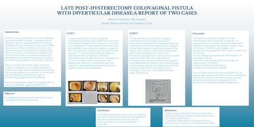
75 year old,P4L4 with history of vaginal hysterectomy 26 years back presented with yellow discharge from vagina for 6 months for which she was treated with multiple doses of antibiotics. She got referred to gyne team by colorectal surgeons when on speculum examination feculent matter was seen. CT scan showed collection lateral to sigmoid with fistula leading to left fornix .Sigmoidoscopy showed stricture at rectosigmoid junction with few diverticulae. Patient had laparoscopic Hartmann’s procedure with end colostomy. .Histopathology showed diverticular disease confirming the diagnosis
Case 2
70 year old P4L4 with a history of vaginal hysterectomy 19 years back and known diverticulosis presented with passage of feculent material for a long time. Speculum examination showed discharge coming from the apex of the vagina .MRI was done which showed extensive sigmoid diverticulosis with colonic wall thickening and sigmoid in close proximity to the apex and confirmed colo-vaginal fistula. .Intraoperatively, sigmoid was densely adherent to the left adnexa and fistula was noted draining into the vagina. Fistula was taken down and had colectomy with end to end anastomosis. Histology in this case confirmed diverticular origin of the fistula
Discussion
Intraabdominal fistulas may be traumatic, inflammatory ,post operative complication or subsequent to a neoplasm. Both the cases had a background of diverticular disease, though it was not known in the first case to begin with. Diverticulosis is a disease of the modern world and is on the rise. A triad of following should raise the suspicion of colovaginal fistula: 1.Recurrent yellowish/purulent discharge not responding to antibiotics 2.A prior history of hysterectomy 3.A history of diverticular disease In such cases, sigmoid becomes adherent to the vaginal apex due to prior inflammation or adhesions. Uterus with its thick myometrial layers may be a preventive factor which predisposes to the formation of these fistulas after hysterectomy
Conclusion
Colovaginal fistulas are uncommon but not rare. We recommend that patients be managed as a part of multidisciplinary team in order to optimize patient care and avoid delays in diagnosis
References
1.Laparoscopic treatment of post-hysterectomy colovaginal fistula in diverticular disease. Minerva Chir 2004 Jun;59(3):301-5. 2. Late Post-Hysterectomy Colovaginal Fistula with Extensive Diverticular Disease October 2020