296 posters, 7 videos, 13 audios, 14 topics, 10 sessions, 1,019 authors, 259 institutions
ePostersLive by SciGen Technologies S.A. All rights reserved.
RCOphth Annual Congress 2026
18 - 21 May, 2026 | Manchester Central, Manchester

P056
The Vanishing Drusen- A Deceptive Case of Primary Intraocular Lymphoma
Poster Presenter
Authors
Part of Topic
Case reports
PURPOSE
Primary intraocular lymphoma (PIOL) is widely regarded as the most deceptive masquerade syndrome in ophthalmology (1-2). We report a case of Diffuse Large B-Cell Lymphoma (DLBCL) that successfully imitated the natural history of bilateral dry age- related macular degeneration before revealing itself through subretinal infiltrates, vitritis, and ultimately CNS involvement. This case highlights the diagnostic pitfalls of PIOL as a masquerade syndrome, the limitations of vitreous biopsy, and the critical importance of maintaining a high index of suspicion in atypical posterior segment presentations.
INTRODUCTION
Primary intraocular lymphoma is a rare, aggressive subset of primary central nervous system lymphoma (PCNSL), most commonly presenting as diffuse large B-cell lymphoma (3) Diagnosis is notoriously delayed, with a reported misdiagnosis rate of 64% and a mean delay from symptom onset to diagnosis of 10 months (4-5).
The clinical significance of PIOL lies in its strong association with CNS lymphoma: approximately 42–92% of patients with PIOL will develop PCNSL within a mean of 8–29 months (6). Early recognition is therefore critical, as CNS involvement carries a substantially worse prognosis, with a 5-year overall survival of approximately 70%.
CASE PRESENTATION
Week 0 — Presentation & Misdiagnosis
Progressive LE central scotoma; slow onset over several months, good navigation preserved
Bilateral RPE changes, scattered drusen, right macular atrophy
Dx: Bilateral dry AMD
Week 6 — First Red Flags
Dramatic drusenoid PED resolution — noted as atypical New peripheral white lesion LE — absent on all prior photographs
Week 10 — Escalating Suspicion
VA 6/6 OU; persistent LE central blur
Marked bilateral subretinal/RPE infiltrates + mild vitritis OU Orbit MRI: non-diagnostic (occupational artefact)
Dx revised: Intermediate uveitis — PIOL suspected
Week 14 — Diagnostic Vitrectomy & Complication
Left vitreous biopsy performed
Superior retinal tear — SF6 tamponade + endolaser required Cytology: inconclusive — abnormal cells, B-cell unconfirmed
Month 4-6 — Therapeutic Stalemate
Prednisolone 40mg + IOP management (timolol/dorzolamide) Haematology: unable to treat — subtype unconfirmed Steroids stopped; patient consents to RE biopsy; referred to Moorfields
Month 9 — Systemic Progression
"lumpy-bumpy" subretinal deposits LE on OCT
Rapid confusion + gait deterioration over days
PET-CT: 2.5cm FDG-avid periventricular mass — absent on MRI just 14 weeks prior
Dx confirmed: DLBCL — CNS + ocular involvement
Month 12— Treatment & Outcome
MATRIX protocol commenced (MTX · AraC · Thiotepa · Rituximab) + stem cell transplant
Eyes stable on monthly review
6/6 RE , 6/36 improved to 6/9 with pinhole LE
Ongoing systemic therapy · 6-monthly ophthalmic surveillance
THE THREE DIAGNOSTIC RED FLAGS:
ThIs case highlighted hree red flags that, in retrospect, pointed strongly toward lymphoma rather than benign macular disease:
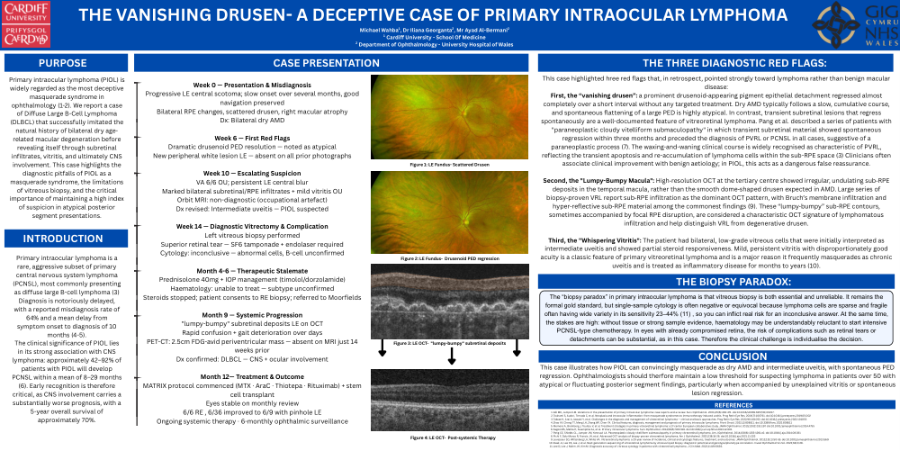
First, the “vanishing drusen”: a prominent drusenoid‐appearing pigment epithelial detachment regressed almost completely over a short interval without any targeted treatment. Dry AMD typically follows a slow, cumulative course, and spontaneous flattening of a large PED is highly atypical. In contrast, transient subretinal lesions that regress spontaneously are a well-documented feature of vitreoretinal lymphoma. Pang et al. described a series of patients with "paraneoplastic cloudy vitelliform submaculopathy" in which transient subretinal material showed spontaneous regression within three months and preceded the diagnosis of PVRL or PCNSL in all cases, suggestive of a paraneoplastic process (7). The waxing-and-waning clinical course is widely recognised as characteristic of PVRL, reflecting the transient apoptosis and re-accumulation of lymphoma cells within the sub-RPE space (3) Clinicians often associate clinical improvement with benign aetiology; in PIOL, this acts as a dangerous false reassurance.
Second, the "Lumpy-Bumpy Macula": High‐resolution OCT at the tertiary centre showed irregular, undulating sub‐RPE deposits in the temporal macula, rather than the smooth dome‐shaped drusen expected in AMD. Large series of biopsy‐proven VRL report sub‐RPE infiltration as the dominant OCT pattern, with Bruch’s membrane infiltration and hyper‐reflective sub‐RPE material among the commonest findings (9). These “lumpy‐bumpy” sub‐RPE contours, sometimes accompanied by focal RPE disruption, are considered a characteristic OCT signature of lymphomatous infiltration and help distinguish VRL from degenerative drusen.
Third, the "Whispering Vitritis": The patient had bilateral, low-grade vitreous cells that were initially interpreted as intermediate uveitis and showed partial steroid responsiveness. Mild, persistent vitritis with disproportionately good acuity is a classic feature of primary vitreoretinal lymphoma and is a major reason it frequently masquerades as chronic uveitis and is treated as inflammatory disease for months to years (10).
THE BIOPSY PARADOX:
The “biopsy paradox” in primary intraocular lymphoma is that vitreous biopsy is both essential and unreliable. It remains the formal gold standard, but single‐sample cytology is often negative or equivocal because lymphoma cells are sparse and fragile often having wide variety in its sensitivity 23–44% (11) , so you can inflict real risk for an inconclusive answer. At the same time, the stakes are high: without tissue or strong sample evidence, haematology may be understandably reluctant to start intensive PCNSL‐type chemotherapy. In eyes with already compromised retina, the risk of complications such as retinal tears or detachments can be substantial, as in this case. Therefore the clinical challenge is individualise the decision.
CONCLUSION
This case illustrates how PIOL can convincingly masquerade as dry AMD and intermediate uveitis, with spontaneous PED regression. Ophthalmologists should therfore maintain a low threshold for suspecting lymphoma in patients over 50 with atypical or fluctuating posterior segment findings, particularly when accompanied by unexplained vitritis or spontaneous lesion regression.