296 posters, 7 videos, 13 audios, 14 topics, 10 sessions, 1,019 authors, 259 institutions
ePostersLive by SciGen Technologies S.A. All rights reserved.
RCOphth Annual Congress 2026
18 - 21 May, 2026 | Manchester Central, Manchester

P038
Diabetic Retinopathy Masking NAION in a Patient on Semaglutide: A Case of Diagnostic Challenge.
Part of Topic
Case reports
Title: Diabetic Retinopathy Masking NAION in a Patient on Semaglutide: A Case of Diagnostic Challenge
Authors: Roque De Monte Furtado¹, Yathavi Charavanamuttu¹, Miss Vivi Riga²
Affiliations: ¹GKT School of Medical Education, King's College London, London, UK; ²Department of Ophthalmology, Guy's and St Thomas' Hospital NHS Foundation Trust, London, UK
Background
-
Non-arteritic anterior ischaemic optic neuropathy (NAION) is characterised by acute painless visual loss, relative afferent pupillary defect, impaired colour vision, ischaemic sectoral optic disc oedema and visual field defects.
-
It is thought to be a result of acute ischaemia of the optic nerve head, though the exact pathogenesis remains unclear.
-
Reports suggest an association between NAION and glucagon-like peptide-1 receptor agonists (GLP-1 RAs).
-
Here we present a case of NAION in a diabetic patient on semaglutide that was initially misdiagnosed as non-ischaemic central retinal vein occlusion (CRVO).
Case Details
-
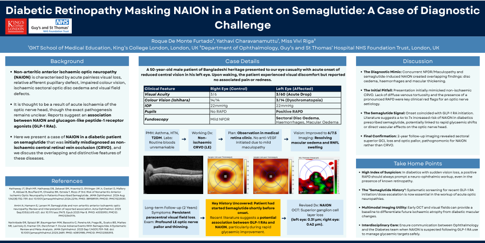
Patient: 50-year-old male of Bangladeshi heritage with acute onset of reduced central vision in his left eye.
-
Medical History: Asthma, HTN, T2DM. Routine bloods unremarkable.
-
Clinical Features (Left Eye):
-
Visual Acuity: 3/60 (Acute Drop)
-
Colour Vision (Ishihara): 3/14 (Dyschromatopsia)
-
IOP: 22mmHg
-
Pupils: Positive RAPD
-
Fundoscopy: Sectoral Disc Oedema, Haemorrhages, Macular Oedema
-
-
Initial Working Dx: Non-ischaemic CRVO (LE).
-
Follow-up: Vision improved to 6/7.5; Imaging showed resolving macular oedema and RNFL swelling.
-
Key History Uncovered: Patient had started Semaglutide shortly before onset.
-
Revised Dx: NAION based on superior ganglion cell layer loss (LE: 0.31 µm; RE: 0.42 µm).
Discussion
-
The Diagnostic Mimic: Concurrent NPDR/Maculopathy and semaglutide-induced NAION created overlapping findings.
-
The Initial Pitfall: Presentation mimicked CRVO, but the presence of a pronounced RAPD and lack of venous tortuosity were key red flags for an optic nerve aetiology.
-
The Semaglutide Signal: Literature suggests a 4x to 7x increased risk of NAION in diabetics prescribed semaglutide.
-
Final Confirmation: 2-year follow-up imaging revealed sectoral superior GCL loss and optic pallor, pathognomonic for NAION.
Take Home Points
-
High Index of Suspicion: In diabetics with sudden vision loss, a positive RAPD should prompt a neuro-ophthalmic workup regardless of retinopathy.
-
The "Semaglutide History": Screening for recent GLP-1 RA initiation is essential in acute optic neuropathy workups.
-
Multimodal Imaging Utility: Early OCT and visual fields help differentiate ischaemic atrophy from diabetic macular changes.
-
Interdisciplinary Care: Ensure communication between Ophthalmology and the Diabetes team to manage glycaemic targets safely.
References
-
Hathaway JT, et al. JAMA Ophthalmol. 2024 Aug 1;142(8):752-759.
-
Amini A, et al. Acta Ophthalmol. 2025 Sep;103(6):615-621.
-
Natividade GR, et al. JAMA Ophthalmol. 2025 Sep 1;143(9):759-768.