Loading...

296 posters, 7 videos, 13 audios, 14 topics, 10 sessions, 1,019 authors, 259 institutions

ePostersLive by SciGen Technologies S.A. All rights reserved.

RCOphth Annual Congress 2026

18 - 21 May, 2026 | Manchester Central, Manchester

RCOphth Annual Congress 2026 banner

P294

Functional and Structural outcome of Amniotic Membrane Graft for repair of Posterior Scleral Perforation with intraocular foreign body. Results from 2 cases
Part of Topic

Vitrea retinal diseases and surgery

Video

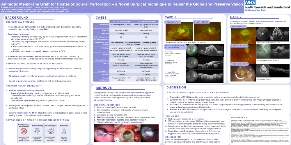
Amniotic Membrane Graft for Posterior Scleral Perforation – a Novel Surgical Technique to Repair the Globe and Preserve Vision

Authors: Thomas W. McNally, Matthew L. Barker, Jonathan Smith, Maged Habib

Lead author and operating surgeon: Maged Habib MSc Ophth FRCS FRCOphth MD

Sunderland Eye Infirmary, South Tyneside and Sunderland NHS Foundation Trust

Background:

THE CLINICAL PROBLEM

•Posterior scleral perforation: rare but devastating open-globe injury classically caused by high-velocity foreign bodies (FBs)
•Poor visual prognosis:
•Posterior perforating wounds carry a poor visual prognosis with ≥40% of patients left with a final visual acuity of NPL/PL[1]
•Outcomes vary depending on mechanism, location and other patient/injury related factors[1,2]:
•Retinal detachment in 70-90% of cases, proliferative vitreoretinopathy in 66% of cases
•Phthisis, enucleation or need for keratoprosthesis in 56%
•Anatomically inaccessible: wounds posterior to the equator are obscured by extraocular muscles (EOMs) and orbital fat making direct external repair infeasible

PRIMARY SURGICAL REPAIR WITHIN 24 HOURS[3]

•Wound exploration: involving conjunctival peritomy + identification of posterior extensions of wounds
•Ab-externo repair: for anterior wounds, working from anterior to posterior
•Occult or posterior wounds: challenging with limited repair options

FURTHER REPAIR METHODS[1-4]

•If left to heal by secondary intention: 
•Loss of globe integrity: leading to hypotony and phthisis bulbi
•Tissue incarceration: high risk for proliferative vitreoretinopathy and retinal detachment
•Sympathetic ophthalmia: higher risk if globe is not closed
•Autologous Tenon plugs: limited to smaller defects, fragile, prone to dislodgement, no antifibrotic effect
•Donor sclera/tenons +/- fibrin glue: tissue availability restricted; donor sclera is rigid, leading to poor conformation to globe curvature

Methods:

We report the results of ab-interno amniotic membrane graft for posterior scleral perforation in two cases of severe penetrating trauma. Both cases were operated by Mr M Habib, consultant vitreoretinal surgeon.

SURGICAL TECHNIQUE

1.Anterior scleral perforation closed primarily
2.25g pars plana vitrectomy with careful vitrectomy around perforation site
3.Endolaser around perforation site
4.AMG introduced via trochar: bimanual under direct observation
5.Amniotic membrane graft to plug posterior wound
6.Silicone oil tamponade for prolonged scleral support

Discussion:

EVIDENCE BASE – ophthalmic use of AMG established

•Zhang Q et al.[6]: AMG used to close a posterior scleral perforation and choroidal hole (case report)
•Suwandi J et al.[7]: “internal plug" technique using two large sheets of amniotic membrane can effectively repair extensive posterior scleral perforations without donor tissue
•Ma D et al.[8]: Amniotic membrane grafting is a viable surgical option for managing severe scleral melting and corneoscleral ulcers, achieving complete re-epithelialisation.
•Hanada K et al.[9]: Multi-layered transplantation acts as a biological scaffold to fill stromal defects, effectively restoring deep corneal and scleral ulcers.

Our cases:

•Globe integrity preserved at >7 months
•IOP normalised in both cases: AMG provides a watertight seal sufficient to maintain globe pressure during scleral remodeling
•VA outcomes confounded: submacular haemorrhage and dense cataract were sequelae of original trauma, not graft failure
•No phthisis, no inflammation  stable globe at 7–9 months supports AM's antifibrotic and immunomodulatory profile

References:


[1]Bleicher ID, Tainsh LT, Gaier ED, Armstrong GW. Outcomes of Zone 3 Open Globe Injuries by Wound Extent: Subcategorization of Zone 3 Injuries Segregates Visual and Anatomic Outcomes. Ophthalmology. 2023 Apr;130(4):379-386.

[2] Dave VP, Das AV, Nayak S, Panchal B, Kuhn F. Clinical Presentations, Management, and Factors Affecting Outcomes in Posterior Segment Open Globe Injuries: An Analysis of 2,360 Eyes. Ophthalmologica. 2023;246(2):150-157.

[3] Blanch RJ, McMaster D, Patterson TJ. Management of open globe injury: a narrative review. Eye (Lond). 2024 Nov;38(16):3047-3051. doi: 10.1038/s41433-024-03246-3.

[4] Zhou Y, DiSclafani M, Jeang L, Shah AA. Open Globe Injuries: Review of Evaluation, Management, and Surgical Pearls. Clin Ophthalmol. 2022 Aug 10;16:2545-2559

[5] Kuhn F, Maisiak R, Mann L, Mester V, Morris R, Witherspoon CD. The Ocular Trauma Score (OTS). Ophthalmol Clin North Am. 2002 Jun;15(2):163-5, vi

[6] Zhang Q, Xia X, Xiang H, Xiong S. Transplantation of amniotic membrane for posterior scleral perforation combined with choroidal hole: A case report. Asian J Surg. 2024 Mar;47(3):1544-1545.

[7] Ma DH, Wang SF, Su WY, Tsai RJ. Amniotic membrane graft for the management of scleral melting and corneal perforation in recalcitrant infectious scleral and corneoscleral ulcers. Cornea. 2002 Apr;21(3):275-83.

[8]Hanada K, Shimazaki J, Shimmura S, Tsubota K. Multilayered amniotic membrane transplantation for severe ulceration of the cornea and sclera. Am J Ophthalmol. 2001 Mar;131(3):324-31.

[9] Suwandi JS, van der Wel BA, van Dijk EHC, Manning SS, Manzulli M, Geeraert P, van Romunde S, Faridpooya K. Internal closure of a posterior perforation using amniotic membrane: A novel surgical technique. Acta Ophthalmol. 2026 Mar;104(2):236-238.


rco2026 banner
Copyright © 2015-2026, ePostersLive® SciGen is a registered trademark of SciGen Technologies S.A. Patent pending.

This website and third-party tools we use rely on cookies for the best user experience. By selecting "I agree", you agree to cookie usage as described in our Privacy Policy.