296 posters, 7 videos, 13 audios, 14 topics, 10 sessions, 1,019 authors, 260 institutions
ePostersLive by SciGen Technologies S.A. All rights reserved.
RCOphth Annual Congress 2026
18 - 21 May, 2026 | Manchester Central, Manchester

P173
Mid-Peripheral Preservation with Temporal Macular and Peripheral Thinning on Widefield OCTA: A Novel Spatial Pattern of Retinal Ischemia in Sickle Cell Retinopathy?
Poster Presenter
Part of Topic
Medical retina
Mid-Peripheral Preservation with Temporal Macular and Peripheral Thinning on Widefield OCTA: A Novel Spatial Pattern of Retinal Ischaemia in Sickle Cell Retinopathy?
Dr. Ikhlas Mahmoud, Dr. Loredana Smarandache, Mr. James Talks Ophthalmology Department, Royal Victoria Infirmary, Newcastle upon Tyne, United Kingdom
PURPOSE
To describe a distinct spatial pattern of retinal thinning on wide field optical coherence tomography angiography (WF-OCTA) thickness maps in sickle cell retinopathy (SCR), defined by a preserved mid-peripheral region between areas of temporal macular and peripheral thinning.
SETTINGS
Ophthalmology Department, Royal Victoria Infirmary, Newcastle upon Tyne, United Kingdom
METHODS
As part of routine SCR monitoring, patients underwent ultra-widefield colour imaging (OPTOS), macular OCT/OCTA (Heilderberg Spectralis), and WF-OCTA (Canon). Thickness maps from 11 patients (22 eyes) were retrospectively reviewed. During this review, a recurring pattern was noted in which temporal macular thinning appeared separated from more peripheral thinning by a mid-peripheral region of relatively preserved thickness.
RESULTS
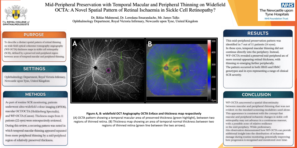
This mid-peripheral preservation pattern was identified in 7 out of 11 patients (14 eyes). In these eyes, temporal macular thinning did not continue directly into the periphery. Instead, WF-OCTA revealed a preserved mid-peripheral arc of more normal-appearing retinal thickness, with thinning re-emerging farther peripherally. The pattern occurred in both HbSS and HbSC genotypes and in eyes representing a range of clinical SCR severity.
CONCLUSION
WF-OCTA uncovered a spatial discontinuity between macular and peripheral thinning that was not evident on the standard screening modalities used alone. This appearance is consistent with the concept that macular and peripheral ischaemic changes in sickle cell retinopathy may not advance in a continuous manner, with a possible zone of relative resilience in the mid-periphery. While preliminary, this observation demonstrated how WF-OCTA can provide additional insight into the distribution of ischaemic damage during routine monitoring, potentially improving how progression is recognised and monitored over time.
Figure A, B: widefield OCT Angiography OCTA Enface and thickness map respectively (A) OCTA pattern showing a temporal macular area of preserved thickness (green highlight), between two regions of thinned retina. (B) Thickness map showing an area of temporal normal thickness between two regions of thinned retina (green line between the two arrows).