296 posters, 7 videos, 13 audios, 14 topics, 10 sessions, 1,019 authors, 260 institutions
ePostersLive by SciGen Technologies S.A. All rights reserved.
RCOphth Annual Congress 2026
18 - 21 May, 2026 | Manchester Central, Manchester

P164
From Retinitis Pigmentosa to Joubert Syndrome: The Impact of Genomic Testing in Retinal Dystrophy Diagnosis
Part of Topic
Medical retina
From Retinitis Pigmentosa to Joubert Syndrome: The Impact of Genomic Testing in Retinal Dystrophy Diagnosis
Introduction
Inherited retinal dystrophies (IRDs) are a heterogeneous group of progressive disorders with variable onset and systemic associations1. Retinitis pigmentosa (RP) is the most common phenotype2, but syndromic ciliopathies such as Joubert syndrome (JS) may present with similar retinal findings3,4. JS is characterised by cerebellar and brainstem malformations, developmental delay, hypotonia, and oculomotor abnormalities3,4. Variants in AHI1 are strongly associated with JS type 3 and retinal degeneration5–7.
History
A patient presented with progressive visual field constriction and long-standing visual impairment, initially suspected as RP. Congenital oculomotor apraxia raised suspicion of a syndromic ciliopathy 3,4.
Examination
Reduced visual acuity, concentric peripheral field loss, bone-spicule pigmentation, vascular attenuation, optic disc pallor, and relative macular preservation.
Investigations
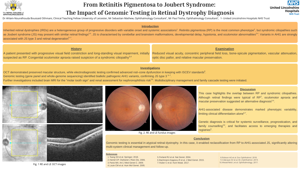
OCT demonstrated preserved macular structure, while electrodiagnostic testing confirmed advanced rod–cone dysfunction in keeping with ISCEV standards8 . Genomic testing (gene panel and whole-genome sequencing) identified biallelic pathogenic AHI1 variants, confirming JS type 3 5–7 . Further investigations included brain MRI for the “molar tooth sign” and renal assessment for nephronophthisis risk . Multidisciplinary management and family cascade testing were initiated.
Discussion
This case highlights the overlap between RP and syndromic ciliopathies. Although retinal findings were typical of RP2 , oculomotor apraxia and macular preservation suggested an alternative diagnosis 3,4 . AHI1-associated disease demonstrates marked phenotypic variability, limiting clinical differentiation alone6,7 . Genetic diagnosis is critical for systemic surveillance, prognostication, and family counselling 3,6, and facilitates access to emerging therapies and registries9.
Conclusion
Genomic testing is essential in atypical retinal dystrophy. In this case, it enabled reclassification from RP to AHI1-associated JS, significantly altering multi-system clinical management and follow-up.