58 posters, 14 topics, 4 sessions, 188 authors, 62 institutions
ePostersLive by SciGen Technologies S.A. All rights reserved.
PSRC 69th Annual Meeting 2024
May 16-19, 2024 | The Westin Copley Place, Boston, MA

EP50
Mri Evaluation Of Neuromas In Oncological Amputees
Part of Topic
Nerve
Introduction
Tumors in the extremities often require amputation for oncologic cure.1 Symptomatic neuroma following amputation are a primary cause of neuropathic pain. MRI features of symptomatic neuromas have been described previously, and radiographic geometrical features did not show difference between symptomatic and non-symptomatic neuromas.2,3 However, it remains unknown to what extend time affects neuroma growth and whether morphology influences pain intensity. Therefore, we aim to assess whether time influences neuroma size and shape, and their association with pain intensity in oncological amputees.
Methods
Oncological patients who underwent extremity amputation were included, MRIs were assessed post-amputation and prior to potential neuroma surgery. Pain of the residual limb, expressed on a Numeric Rating Scale (NRS, 0-10) within 3 months of the MRI assessment, and presence of neuropathic symptoms, were recorded from chart review.
Neuromas were classified as symptomatic neuroma, or non-symptomatic neuroma, based on description of pain with neuropathic symptoms. Two musculoskeletal radiologists independently assessed the MR images and collected MRI features per neuroma. Discrepancy was solved by a third musculoskeletal radiologist serving as independent adjudicator. Neuroma size was expressed as the neuroma-to-nerve-ratio (NNR).
Results
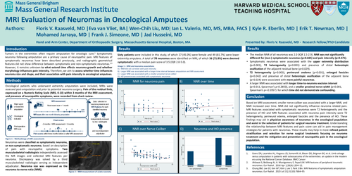
Sixty patients were included in this study, of which 27 (45.0%) were female and 49 (81.7%) were lower extremity amputees. A total of 78 neuromas were identified on MRI, of which 56 (71.8%) were deemed symptomatic with a median pain score of 3.5 (IQR 2.0-5.0).
Conclusion
Based on MRI assessment, smaller nerve caliber was associated with a larger NNR, and NNR increased over time. NNR did not significantly influence neuroma related pain. MRI features associated with symptomatic neuromas were T2 heterogeneity and the presence of HO and MRI features associated with neuroma pain intensity were T2 heterogeneity, perineural edema, enlarged fascicles and the presence of HO. These findings may aid in physician awareness of neuromas in the oncological population and assist in the selection of patients for surgical neuroma treatment. Understanding the relationship between MRI features and pain score can aid in pain management strategies for patients with neuromas. These results may help in more refined patient stratification and selection for nerve surgical treatments focusing on neuroma treatment and the mitigation and prevention of neuropathic pain in the oncological population.
References