58 posters, 14 topics, 4 sessions, 188 authors, 62 institutions
ePostersLive by SciGen Technologies S.A. All rights reserved.
PSRC 69th Annual Meeting 2024
May 16-19, 2024 | The Westin Copley Place, Boston, MA

EP39
3D-Printed Microneedles For Lymphatic Mapping
Poster Presenter
Authors
Affiliations
Part of Topic
Innovation/Commercialization/Technology
3D-Printed Microneedles for Lymphatic Mapping
BACKGROUND
Many patients with extremity lymphedema anticipating lymphaticovenous anastomosis cannot tolerate the pain caused by numerous intradermal contrast injections during lymphatic mapping in the outpatient setting. Performing lymphatic mapping intraoperatively incurs increased costs and morbidity associated with longer anesthesia. The use of microneedles is an attractive alternative that may reduce pain for the patient while also providing sufficient intradermal contrast.
OBJECTIVE
The purpose of this proof-of-concept project was to design, fabricate, and test microneedles tailored for intradermal contrast delivery in the context of lymphatic mapping using both microbubbles for contrast-enhanced ultrasound and indocyanine green (ICG) for near-infrared imaging.
METHODS
Two array configurations (27-microneedle and 3- microneedle), two lumen diameters (100 µm and 200 µm), and two needle lengths (1.1 mm and 1.6 mm) were tested in ex vivo porcine skin, in an animal model, and in cadaveric skin.
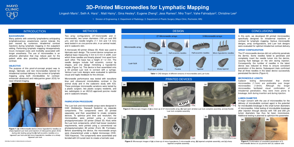
A microscale 3D printer (Dilase 3D, Kloé) was used to fabricate each design. The current device comprises an elliptical base measuring 3 mm to 6 mm in width, over which three needles are linearly placed 1mm apart from each other. The base has a height of 1.2 mm. The needle designs include half eccentric, conical tip shapes, and one design mimicking a hypodermic needle (Figure 1a). This device is then assembled onto a 3D printed Luer lock to connect with a syringe. Various Luer Lock designs were evaluated to optimize visual and haptic feedback for the clinician.
Microneedle performance was tested with isosulfane blue and ultrasound microbubbles contrast in the animal study and in the cadaveric study with ICG. These prototype microneedle variations were tested by a plastic surgeon, two plastic surgery residents, and two radiologists in an IACUC-approved porcine model and in cadaveric breasts.
The Luer lock and microneedle arrays were designed in CAD (Solidworks, Dassault Systems) as separate components. The components were 3D printed separately and then assembled to create complete devices. To optimize print time and resolution, the microneedles were printed using a micro-vat photopolymerization 3D printer (Dilase 3D, Kloé) while the Luer lock components, which had looser resolution requirements, were printed using a conventional vat photopolymerization 3D printer (Form 3B, Formlabs). Before assembling the device, the microneedle arrays were characterized under a digital microscope (VHX-7000 Keyence). The components were assembled and bonded with UV-cured resin to create a hermetic seal.
CONCLUSIONS
In this work, we developed 3D printed microneedles specifically designed for intradermal injections of contrast for lymphatic mapping. Various microneedle designs, array configurations, and Luer lock designs were evaluated for optimal intradermal contrast delivery.
The 27-microneedle devices did not uniformly penetrate the dermis. Some needles failed to penetrate and completely insert the needle lumen into the dermis, causing fluid leakage on the skin during injection. Consequently, the number of needles in the latest device was reduced to three to ensure consistent penetration of the dermis. Subsequent trials confirmed that all three needles in the latest device successfully penetrated the dermis (Figure 4).
Cadaver testing demonstrated that shorter microneedles (1.1mm) are preferable over longer microneedles (1.6mm). Although the longer microneedles facilitated visual confirmation of intradermal penetration, they were more prone to breakage, both during insertion and during injection.
A major concern with the use of microneedles for the delivery of microbubble contrast agent is the potential for microbubble breakage in the small lumen diameters of microneedles. Initial testing of microbubble breakage after injection through devices with 100 and 200 µm lumen diameters has thus far been inconclusive. Additional testing is planned to optimize this parameter.