358 posters, 97 topics, 122 sessions, 958 authors, 239 institutions
ePostersLive by SciGen Technologies S.A. All rights reserved.
79th PostGraduate Assembly in Anesthesiology 2025
December 12 - 15, 2025 | New York City

2112
Management of Acute Opioid-Induced Respiratory Failure in Patient with a Large Mediastinal Mass.
Poster Presenter
Authors
Affiliations
Part of Topic
Airway - Critical Care
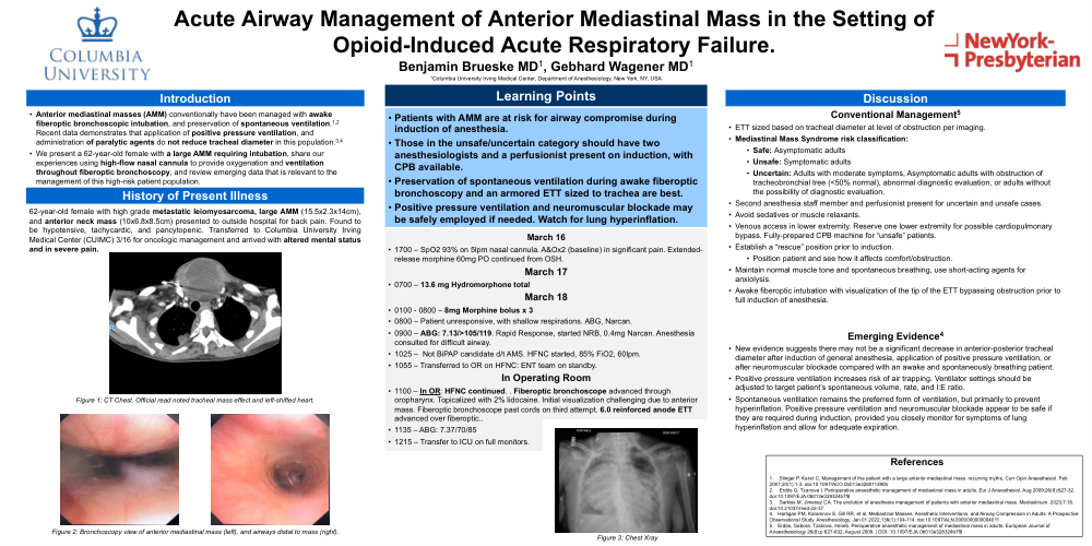
Acute Airway Management of Anterior Mediastinal Mass in the Setting of Opioid-Induced Acute Respiratory Failure
Benjamin Brueske MD¹, Gebhard Wagener MD¹
¹Columbia University Irving Medical Center, Department of Anesthesiology, New York, NY, USA
Introduction
-
Anterior mediastinal masses (AMM) conventionally have been managed with awake fiberoptic bronchoscopic intubation, and preservation of spontaneous ventilation.¹,² Recent data demonstrates that application of positive pressure ventilation, and administration of paralyzing agents do not reduce tracheal diameter in this population.³,⁴
-
We present a 62-year-old female with a large AMM requiring intubation, share our experiences using high-flow nasal cannula to provide oxygenation and ventilation throughout awake fiberoptic bronchoscopy, and review emerging data that is relevant to the management of this high-risk patient population.
History of Present Illness
62-year-old female with high-grade metastatic leiomyosarcoma, large AMM (15.5×2.3×14 cm), and anterior neck mass (10×6.8×8.5 cm) presented to outside hospital for back pain. Found to be hypotensive, tachycardic, and pancytopenic. Transferred to Columbia University Irving Medical Center (CUIMC) 3/16 for oncologic management and arrived with altered mental status and in severe pain.
Timeline
March 16
1700 – SpO₂ 93% on 5 L NC. A&O×2 (baseline) in significant pain. Extended-release morphine 60 mg continued from OSH.
March 17
0700 – 13.6 mg hydromorphone total
March 18
0100–0800 – 8mg Morphine bolus ×3
0800 – Patient unresponsive, shallow respirations. ABG, Narcan.
0900 – ABG: 7.13 / >105 / 119. Rapid Response, started NRB, 0.4 mg Narcan.
Anesthesia consulted for difficult airway.
1025 – HFNC started. 85% FiO₂, 60 Lpm.
1055 – Transfer to OR for HFNFC. ENT on standby.
In Operating Room
1100: HFNC continued. Awake fiberoptic bronchoscope advanced through oropharynx. Topicalized with 2% lidocaine. Initial visualization challenging due to anterior mass. Fiberoscope passed over cords on third attempt. 6.0 reinforced ETT placed.
1130 – ABG: 7.37 / 70 / 85
1215 – Patient transferred to ICU on full monitors.
Learning Points
-
Patients with AMM are at risk for airway compromise during induction of anesthesia.
-
Those in the “unsafe or uncertain” category should have two anesthesiologists and a perfusionist ptrdrny on induction, with CPB available.
-
Preservation of spontaneous ventilation during awake fiberoptic bronchoscopy and armored ETT sized to trachea at the area of compression is best.
-
Positive pressure ventilation and neuromuscular blockade may be safely employed if needed. Watch for lung hyperinflation.
Discussion
Conventional Management⁵
-
ETT sized based on tracheal diameter at level of obstruction per imaging.
-
Mediastinal Mass Syndrome risk classification:
-
Safe: asymptomatic adults
-
Unsafe: symptomatic adults
-
Uncertain: adults with moderate symptoms, asymptomatic adults with obstruction (<50% normal), abnormal diagnostic evaluation, or without the possibility of diagnostic evaluation.
-
-
Second anesthesiologist and perfusionist present for uncertain/unsafe cases.
-
Avoid sedatives or muscle relaxants.
-
Venous access in lower extremity. Reserve one lower extremity for possible CPB cannulation. Fully-prepared CPB machine for “unsafe” patients.
-
Establish a “rescue” position prior to induction:
-
Position patient and see how it affects comfort/obstruction.
-
-
Maintain normal muscle tone and spontaneous breathing, use short-acting agents for anxiolysis.
-
Awake fiberoptic intubation with visualization of ETT tip bypassing obstruction prior to induction of anesthesia.
Emerging Evidence⁴
-
New evidence suggests no significant decrease in anterior–posterior tracheal diameter after induction of general anesthesia, positive pressure ventilation, or neuromuscular blockade compared with awake states.
-
Positive pressure ventilation increases risk of air trapping. Ventilator settings should be adjusted to match patient’s spontaneous volume, rate, and I:E ratio.
-
Spontaneous ventilation remains the preferred form of ventilation, but primarily to prevent hyperinflation. Positive pressure ventilation and neuromuscular blockade appear safe if they are required during induction, provided the anesthesiologist carefully observes for lung hyperinflation and allows for adequate expiration.
References:
1. Slinger P, Karsli C. Management of the patient with a large anterior mediastinal mass: recurring myths. Curr Opin
Anaesthesiol. Feb 2007;20(1):1-3. doi:10.1097/ACO.0b013e328011390b
2. Erdös G, Tzanova I. Perioperative anaesthetic management of mediastinal mass in adults. Eur J Anaesthesiol. Aug
2009;26(8):627-32. doi:10.1097/EJA.0b013e328324b7f8
3. Sarkiss M, Jimenez CA. The evolution of anesthesia management of patients with anterior mediastinal mass.
Mediastinum. 2023;7:16. doi:10.21037/med-22-37
4. Hartigan PM, Karamnov S, Gill RR, et al. Mediastinal Masses, Anesthetic Interventions, and Airway Compression
in Adults: A Prospective Observational Study. Anesthesiology. Jan 01 2022;136(1):104-114.
doi:10.1097/ALN.0000000000004011
5. Erdös, Gabora; Tzanova, Ireneb. Perioperative anaesthetic management of mediastinal mass in adults. European
Journal of Anaesthesiology 26(8):p 627-632, August 2009. | DOI: 10.1097/EJA.0b013e328324b7f8