358 posters, 97 topics, 122 sessions, 958 authors, 239 institutions
ePostersLive by SciGen Technologies S.A. All rights reserved.
79th PostGraduate Assembly in Anesthesiology 2025
December 12 - 15, 2025 | New York City

2161
Dislodged Endotracheal Tube: A Case Report
Poster Presenter
Authors
Affiliations
Part of Topic
Airway - Critical Care
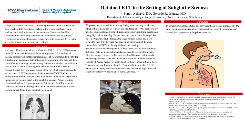
Subglottic stenosis is defined as narrowing from the lower surface of the true vocal cords to the inferior surface of the cricoid cartilage. Causes include congenital or iatrogenic interventions. Treatment should be focused on the underlying condition and maintaining airway patency.1 Tracheostomy tube dislodgement is very rare, with incidence 0.1%. In the event that this occurs, mortality is very high.2 A 42 year old male with a history of anemia, ESRD, HLD, HTN presented to the ED at an outside hospital for throat tightness. CT scan showed tracheal stenosis with soft tissue thickening. Patient was taken to the OR for a tracheotomy and repair. Patient became hypoxic during the case and there was difficulty obtaining a secure airway. Patient presented to our institution with a 4.0 ETT that was dislodged in the right lower lobe, a 5.0 ETT passing through the vocal cords ending at the lip, which was subsequently secured to a 6.0 ETT by our team. Patient received VV ECMO and a bronchoscopy for ETT tube retrieval. Patient was found to have a posterior membrane perforation distal to the subglottic stenosis. Patient was then reintubated with an 8.0 intraoperatively. Afterwards, the CT scan showed decreased mucosal thickening, resolved pneumomediastum, and a healed tracheal defect. Patient was eventually extubated. We present a case of a difficult airway during a tracheotomy repair case, which led to a dislodged ETT. There is a incidence of 1/1000 tracheostomy tube becoming dislodged. While this is a rare occurrence, these events have a very high risk of mortality.3 In our case, our patient had a dislodged 4.0 ETT, a 5.0 uncuffed ETT through the vocal cords at the lip, and a 6.0 secured to the 5.0 ETT. There was concern of perforation of the distal airway from the ETT into the right hilar tissue, causing pneumomediastinum. Management of these cases vary by the respiratory distress of patient, tube position, and acute need to resecure the airway. After the patient is stable, further imaging should be done. Additionally, when using different size tubes to ventilate, Reynold’s number should be considered. With a higher Reynold’s number, there is more turbulent flow. This turbulent gas flow from the 6.0 ET funneled into a smaller 5.0 ET, which is more likely to have laminar flow. The mechanics of gas flow can affect how effectively the patient is being ventilated. 4-5 In conclusion, dislodged tubes can carry significant risk as evidenced by the iatrogenic pneumomediastinum. This patient was promptly identified and treated. Airway patency is the primary concern.