358 posters, 97 topics, 122 sessions, 958 authors, 239 institutions
ePostersLive by SciGen Technologies S.A. All rights reserved.
79th PostGraduate Assembly in Anesthesiology 2025
December 12 - 15, 2025 | New York City

2234
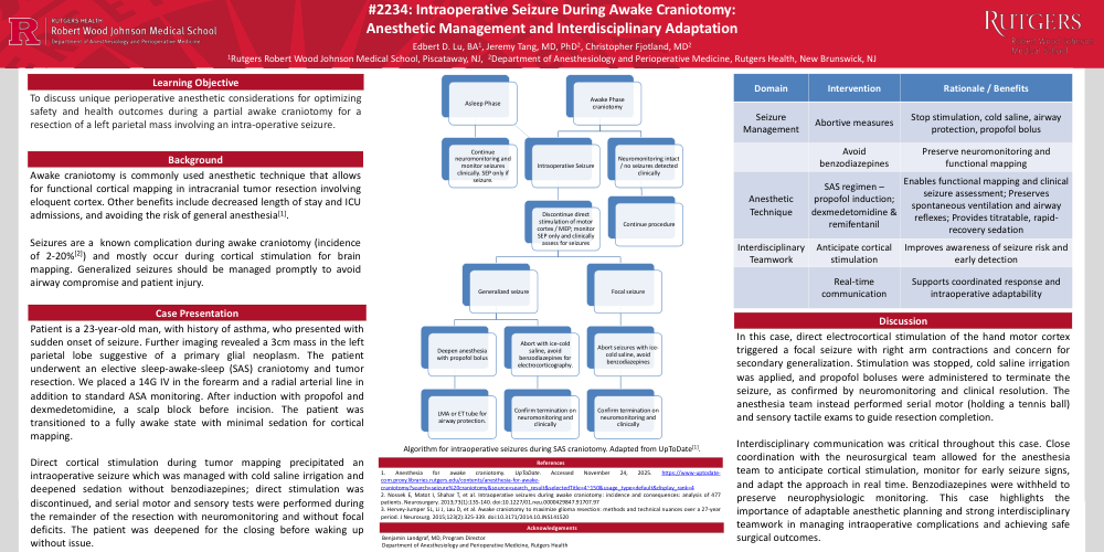
Intraoperative Seizure During Awake Craniotomy: Anesthetic Management and Interdisciplinary Adaptation
Poster Presenter
Authors
Affiliations
Part of Topic
Neuroanesthesia - Perioperative Management
#2234: Intraoperative Seizure During Awake Craniotomy: Anesthetic Management and Interdisciplinary Adaptation
Edbert D. Lu, BA1, Jeremy Tang, MD, PhD2, Christopher Fjotland, MD2
1Rutgers Robert Wood Johnson Medical School, Piscataway, NJ, 2Department of Anesthesiology and Perioperative Medicine, Rutgers Health, New Brunswick, NJ
Learning Objectives
To discuss unique perioperative anesthetic considerations for optimizing safety and health outcomes during a partial awake craniotomy for a resection of a left parietal mass involving an intra-operative seizure.
Background
Awake craniotomy is commonly used anesthetic technique that allows for functional cortical mapping in intracranial tumor resection involving eloquent cortex. Other benefits include decreased length of stay and ICU admissions, and avoiding the risk of general anesthesia[1].
Seizures are a known complication during awake craniotomy (incidence of 2-20%[2]) and mostly occur during cortical stimulation for brain mapping. Generalized seizures should be managed promptly to avoid airway compromise and patient injury.
Case Presentation
Patient is a 23-year-old man, with history of asthma, who presented with sudden onset of seizure. Further imaging revealed a 3cm mass in the left parietal lobe suggestive of a primary glial neoplasm. The patient underwent an elective sleep-awake-sleep (SAS) craniotomy and tumor resection. We placed a 14G IV in the forearm and a radial arterial line in addition to standard ASA monitoring. After induction with propofol and dexmedetomidine, a scalp block before incision. The patient was transitioned to a fully awake state with minimal sedation for cortical mapping.
Direct cortical stimulation during tumor mapping precipitated an intraoperative seizure which was managed with cold saline irrigation and deepened sedation without benzodiazepines; direct stimulation was discontinued, and serial motor and sensory tests were performed during the remainder of the resection with neuromonitoring and without focal deficits. The patient was deepened for the closing before waking up without issue.
Discussion
In this case, direct electrocortical stimulation of the hand motor cortex triggered a focal seizure with right arm contractions and concern for secondary generalization. Stimulation was stopped, cold saline irrigation was applied, and propofol boluses were administered to terminate the seizure, as confirmed by neuromonitoring and clinical resolution. The anesthesia team instead performed serial motor (holding a tennis ball) and sensory tactile exams to guide resection completion.
Interdisciplinary communication was critical throughout this case. Close coordination with the neurosurgical team allowed for the anesthesia team to anticipate cortical stimulation, monitor for early seizure signs, and adapt the approach in real time. Benzodiazepines were withheld to preserve neurophysiologic monitoring. This case highlights the importance of adaptable anesthetic planning and strong interdisciplinary teamwork in managing intraoperative complications and achieving safe surgical outcomes.
1. Anesthesia for awake craniotomy. UpToDate. Accessed November 24, 2025. https://www-uptodate-com.proxy.libraries.rutgers.edu/contents/anesthesia-for-awake-craniotomy?search=seizure%20craniotomy&source=search_result&selectedTitle=4~150&usage_type=default&display_rank=4
2. Nossek E, Matot I, Shahar T, et al. Intraoperative seizures during awake craniotomy: incidence and consequences: analysis of 477 patients. Neurosurgery. 2013;73(1):135-140. doi:10.1227/01.neu.0000429847.91707.97
3. Hervey-Jumper SL, Li J, Lau D, et al. Awake craniotomy to maximize glioma resection: methods and technical nuances over a 27-year period. J Neurosurg. 2015;123(2):325-339. doi:10.3171/2014.10.JNS141520
Acknowledgements
Benjamin Landgraf, MD, Program Director
Department of Anesthesiology and Perioperative Medicine, Rutgers Health